Signet ring cells
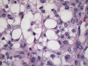
Signet ring cell melanoma is defined by the microscopic appearance of some cells. Cytoplasmic contents displace the nucleus towards the edge, creating a ring-like appearance. It cannot be reliably identified from the colour or shape of a skin lesion.
Descriptions include cells with accumulated filaments, including vimentin, and others with cytoplasmic vacuoles, sometimes resembling lipoblasts. This is not a classification patients can apply to a photograph. It describes morphology rather than origin from gastric carcinoma or a fatty-tissue tumour.
Presentation and lesion origin
This morphology can occur in different settings, including metastatic disease. The pathologist therefore needs to establish whether the sample comes from a primary lesion or a site of spread. Published cases include the trunk and limbs, but these are not mandatory locations.
Case numbers are too limited to establish a reliable universal age pattern or sex preference. A lesion may be nonspecific or poorly pigmented. Clinical assessment follows change, location and personal history rather than trying to recognise signet ring cells externally.
Differential diagnosis and immunohistochemistry
Histological examination assesses the full melanocytic pattern and compares it with other tumours showing similar cells, particularly carcinomas and neoplasms with lipoblast-like features. A history of melanoma and comparison with previous samples are important.
An immunohistochemical panel assesses melanocytic markers and markers of other cellular lineages. Expression can vary: one positive result is insufficient, and one negative result does not exclude every melanoma. Ambiguous cases may benefit from expert review and selected additional investigations.
The report should distinguish morphological description from the final diagnosis and information required for staging. A very limited biopsy can make recognition of different components more difficult.
Treatment and interpretation of prognosis
Rarity and the frequent publication of complex cases do not prove that every melanoma with this morphology has a worse prognosis independently of stage. Small series cannot provide reliable individual estimates.
Surgery is considered for localised disease. Sentinel lymph node biopsy and further investigations depend on the clinical setting; regional lymph node dissection is not automatic. Advanced disease may be considered for immunotherapy, appropriate targeted treatments and other indicated options.
Ask whether the lesion is primary or metastatic, which findings establish stage and whether the sample has undergone expert review. Follow-up planning depends on those answers rather than the variant name alone.
Sources and update
Editorial revision: 17 October 2026. This information does not replace medical assessment. For very rare variants, some evidence comes from small series or case reports.