Myxoid melanoma is a rare variant of cutaneous malignant melanoma that is distinguished by its unique histological appearance. This type of melanoma often manifests as myxoid deposits in melanoma metastases, however primary myxoid melanomas of the skin and sinus have also been described.
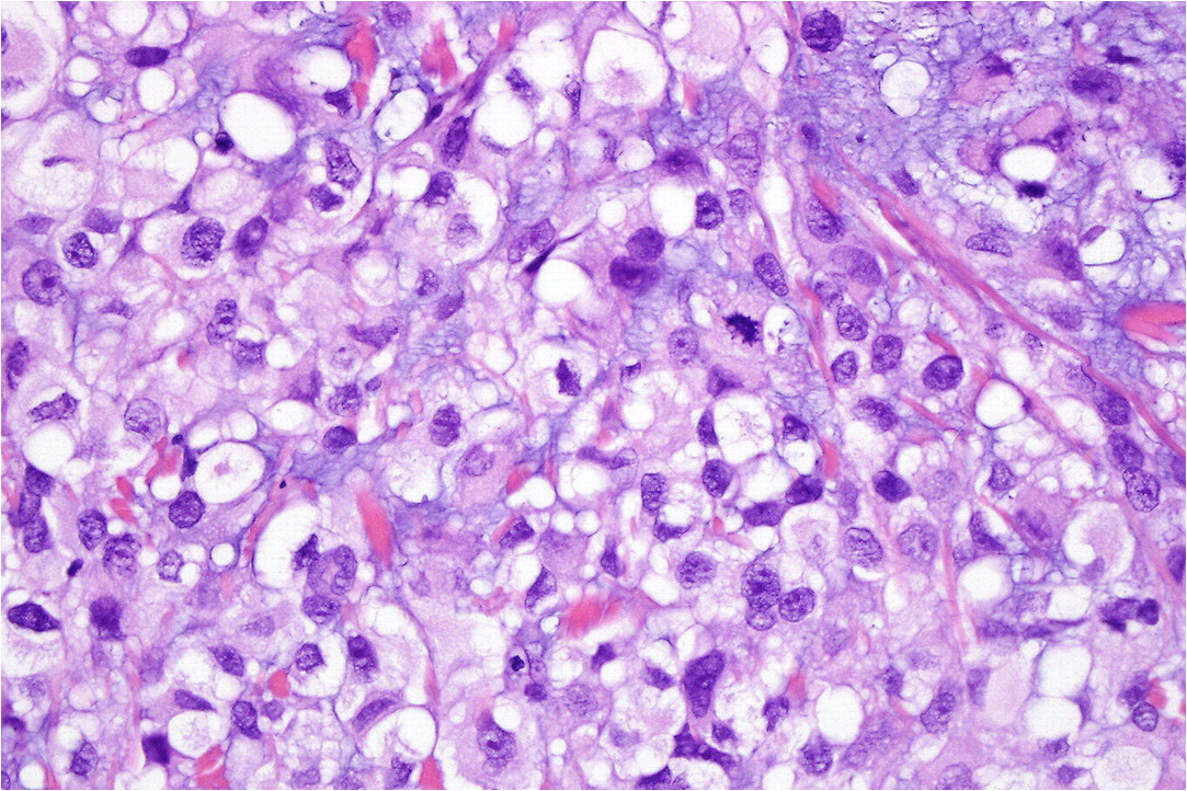
Clinically, myxoid melanoma may present as a rapidly growing skin nodule or mass, located anywhere on the body but most commonly found on the skin of the trunk or extremities. The lesion is characterized by large malignant cells embedded in a myxoid matrix, composed of mucopolysaccharides, which gives the lesion a clear or pale, sometimes cystic or vesicular appearance.
Histologically, tumor cells are usually negative by staining with mucicarmine and PAS diastase, while they are positive by staining with low pH Alcian Blue. The tumor is predominantly amelanotic and the myxoid stroma appears to be produced not by the tumor cells themselves, but by the tissue response to these cells.
The diagnosis of myxoid melanoma may be complicated by its rarity and unusual clinical and histological presentation, which may be confused with other skin lesions. Indicators used for diagnosis include tumor cell positivity for S100 and HMB-45, and an E9 stain may indicate rapid progression.
Treatment of myxoid melanoma generally follows the same approach as for other types of melanoma and may include surgical removal of the lesion, immunotherapy, chemotherapy, or radiation therapy. There are no specific treatment guidelines for myxoid melanoma due to its rarity, therefore each case must be managed on an individual basis.