What having a risk factor means
Melanoma has a multifactorial origin: individual susceptibility, skin characteristics, environmental exposures and medical history can contribute in different ways. A risk factor increases the probability of disease within a group of people; it is not a diagnosis and cannot, on its own, predict an individual’s future. Melanoma can also develop in someone who has no obvious risk factors.
It is useful to distinguish factors we can influence, especially avoidable ultraviolet exposure, from characteristics we cannot change, such as family history, age or inherited susceptibility. Looking at the whole picture helps plan appropriate checks without treating every mole as a cause for alarm. This page mainly concerns cutaneous melanoma: melanomas of the eye and mucosal surfaces have partly different characteristics and predisposing factors.
The child pages explain two tools that are often confused: skin phototypes describe the skin’s response to sunlight, whereas the Glogau classification describes visible photoageing. Neither provides an individual melanoma probability score.
Sunlight, sunburn and UVA and UVB radiation
Ultraviolet exposure is an important environmental risk factor for cutaneous melanoma. Intense, intermittent exposure, such as concentrated holiday exposure, and sunburn are particularly relevant. Burns in childhood and adolescence matter in a person’s risk history, but burns in adulthood also deserve attention. There is no age after which getting sunburnt becomes harmless.
It is incorrect to attribute the problem only to UVB or to regard UVA as safe. Both components of ultraviolet radiation contribute to skin damage; the absence of obvious redness does not demonstrate that an exposure is harmless. Some melanomas are associated with chronic sun damage and others with intermittent exposure: habits, anatomical site and individual susceptibility interact.
Melanoma prevention includes shade, clothing, a hat and protection for uncovered skin. Checking the UV index helps plan outdoor activities: how warm it feels and whether the sky is cloudy are not reliable measures of ultraviolet intensity. Sunscreen is one part of protection, not a way to deliberately extend time in the sun.

Sunlamps and tanning beds
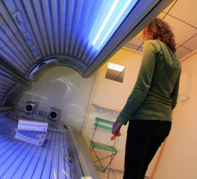
Tanning devices expose the skin to UV radiation and increase skin cancer risk. UV-emitting tanning devices are classified by IARC as carcinogenic to humans. Starting young and accumulating exposures are particularly concerning, but an exposure does not become a problem only after a predetermined number of sessions.
A threshold such as “ten sessions per year” must therefore not be interpreted as a safety limit. An artificial tan does not safely prepare the skin for a holiday and is not recommended as a way to obtain vitamin D. Concerns about deficiency should be discussed with a clinician, without using tanning beds or intentional unprotected exposure.
Phototype and pigmentation: different protection, never zero risk
Skin that burns easily, red or blond hair, light eyes and freckles are associated with greater susceptibility to sun damage and cutaneous melanoma. The Fitzpatrick classification mainly considers the tendency to burn and tan: hair and eye colour help describe a person but do not automatically assign a phototype.
More intense pigmentation provides greater natural protection from UV radiation without conferring immunity to melanoma. Phototype is not the same as nationality, geographical origin or ethnic identity. Population comparisons also depend on behaviour, access to care and cancer registration practices: they are not a personal risk calculator.
Melanoma is less frequent in absolute terms in people with darker skin; acral forms represent a relatively larger share of diagnosed cases. This does not mean that they are the most common cancer in a particular population. Palms, soles and nails deserve attention in every phototype, even though they receive little sunlight.
Number of moles and atypical moles
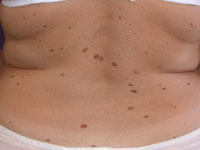
Having many melanocytic moles and atypical moles is a marker of increased risk. A dermatologist considers their number, distribution, appearance and changes over time together with personal history. There is no count that sharply separates a “safe” person from someone destined to develop melanoma, and not every mole has the same clinical significance.
A dysplastic nevus is not synonymous with melanoma. It may indicate susceptibility affecting the whole skin, while most melanomas develop without a recognisable pre-existing mole. Preventively removing all moles therefore does not eliminate risk and is not a general prevention strategy. Suspicious lesions, however, need assessment and, when indicated, removal for histological examination.
For people with many moles, comparison over time, dermoscopy and, in selected cases, photographic documentation can help. Photographs do not replace examination: a new change or a lesion unlike the others needs assessment even if the next scheduled appointment is some time away.
Large and giant congenital moles
Congenital moles require an assessment distinct from that of common acquired moles. Risk varies particularly with projected adult size, the number of associated lesions and certain clinical features. Small and medium lesions carry a much lower risk than large and giant forms; applying one percentage to all lesions, or a fixed 15-centimetre threshold, is misleading.
In more extensive forms, melanoma may occur early and develop deep within the tissues rather than necessarily beginning as a superficial mark. A new nodule that grows, becomes firm, ulcerates or causes persistent symptoms needs specialist attention. Care for children with large, giant or multiple congenital moles should be individualised, also considering possible involvement beyond the skin.
Surgical decisions depend on several goals, including function, appearance and the ability to monitor the lesion. Removing the visible portion does not guarantee elimination of all risk. A shared plan with an experienced centre is helpful, without automatically applying estimates from very different clinical populations to an individual child.
Personal history, family history and genetic counselling
Someone who has already had melanoma has an increased risk of another melanoma: follow-up concerns both the treated disease and the rest of the skin. Melanoma in relatives, especially close relatives, also matters. The number of affected family members, age at diagnosis, multiple melanomas in one person and other cancers in the family all contribute to assessment.
Family history does not mean certainty of developing disease. It is inappropriate to claim that some people with atypical moles and a family history will inevitably develop melanoma during their lifetime. Risk depends on the particular susceptibility and exposures; it cannot be calculated by adding or multiplying the relative risks of different factors.
Recognised susceptibility genes include CDKN2A and, more rarely, CDK4; other predispositions include those associated with BAP1, MITF and POT1. Certain MC1R variants influence pigmentation and susceptibility but do not, on their own, amount to a high-risk inherited syndrome. A variant found in a tumour is not necessarily inherited: tumour testing and constitutional testing answer different questions.
Several melanomas within a family or in one person, early diagnoses and combinations with other cancers may justify genetic counselling. For example, a family combination of melanoma and pancreatic cancer can be relevant. A specialist assesses the indications, limitations and consequences of testing; a negative result does not automatically cancel a significant family history.
Immunosuppression and rare genetic conditions
Organ transplant recipients taking immunosuppressive treatment have an increased risk of skin cancers; the increase is particularly marked for squamous cell carcinoma but also concerns melanoma. Certain blood disorders, such as chronic lymphocytic leukaemia and some lymphomas, may be associated with higher risk. Its magnitude and management depend on the clinical situation: applying the same multiplier to everyone is inappropriate.
These conditions call for surveillance agreed between specialists and attention to sun protection. Immunosuppressive medicines must not be stopped or changed independently to reduce skin cancer risk: their benefits and any adjustments require assessment by the treating team.
Xeroderma pigmentosum is a rare disorder of DNA repair mechanisms, with marked susceptibility to skin cancers and a need for specialist protection and surveillance. Certain inherited cancer syndromes, including hereditary retinoblastoma and Li-Fraumeni syndrome, also require dedicated care pathways. Associations reported for other conditions, including neurofibromatosis, should not be presented as equivalent in evidence strength or absolute risk: the specific clinical picture guides counselling.
Age, sex and interpreting the data
Melanoma can occur at different ages but is mainly a disease of adulthood and is rare in children. Frequency and distribution by sex vary with age and the population being studied. A single average age or survival figures for a narrow age band do not describe the risk of the individual reader.
Incidence, relative risk and survival are different measures: the first describes how many new cases occur, the second compares groups, and the third concerns people who have already been diagnosed. The melanoma epidemiology page places population figures in context. For an individual patient, medical history, lesion characteristics and, after diagnosis, disease stage are what matter.
From risk factors to practical choices
Bring information about personal or family melanomas, previous removals and their pathology reports, significant sunburns, tanning-bed use and current treatments to your appointment. This helps the clinician decide the timing and methods of checks. One schedule for everyone cannot replace that assessment; nor can an app or online questionnaire make a diagnosis.
Daily UV protection and skin self-examination have complementary roles. For uncovered skin, recommendations include broad-spectrum, water-resistant sunscreen with SPF at least 30, applied properly and reapplied as directed, together with shade and clothing. The ABCDE rule and comparison with other lesions help identify changes, without waiting for every criterion to be present.
A changing mole, a new lesion unlike the others, a growing nodule or unexplained bleeding warrants timely assessment. Prevention and early detection are not the same: the former aims to reduce avoidable exposures, the latter to recognise disease at an early stage. No measure eliminates risk, but an understandable care pathway helps ensure that warning signs are not overlooked.
Sources and update
- NCI — Skin Cancer Prevention (PDQ)
- NCI — Common Moles, Dysplastic Nevi, and Risk of Melanoma
- NCI — Genetics of Skin Cancer (PDQ)
- WHO — Ultraviolet radiation
- WHO — Artificial tanning devices
- DermNet — Congenital melanocytic naevi
- American Academy of Dermatology — Sunscreen FAQs
Editorial revision: 17 October 2026. This material is educational and does not replace medical assessment.