Lentigo maligna and invasive melanoma are not synonyms
Lentigo maligna is a form of melanoma in situ: tumour cells are confined to the epidermis and may extend along skin appendages. Once the dermis is invaded, it is called lentigo maligna melanoma. This distinction is essential to interpreting the report, risk and treatment.
It is closely associated with chronically sun-damaged skin and is seen mainly in older people, often on the face and neck. Other sun-exposed sites can also be affected. It should not be confused with a benign solar lentigo simply because both may look like brown patches.
The in situ phase may evolve slowly over years and reach several centimetres. Neither slow growth nor size excludes an invasive component. A suspicious lesion should be assessed before it becomes raised.
Appearance of the patch and changes
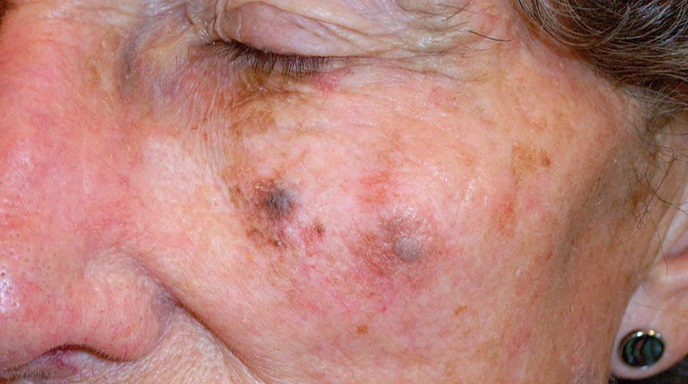
The patch may be asymmetric, with irregular edges and uneven pigmentation. Light brown, dark brown, black and grey may coexist, and some areas can lighten. Its true extent may be difficult to see against already sun-damaged skin.
A new nodule, firmer area, ulceration or bleeding deserves particular attention. These may suggest invasion, but microscopic examination confirms it. Their absence does not prove that the lesion is only in situ.
A history of changes and earlier photographs are useful. Observation includes a familiar patch that changes; variations should not automatically be attributed to age or sunlight.
Dermoscopy and biopsy planning
Dermoscopy can reveal grey dots and globules, asymmetric pigment around follicular openings and irregular structures. These findings must be interpreted together rather than used for self-diagnosis. In specialist centres, reflectance confocal microscopy may assist assessment of selected cases.
Confirmation requires histological examination. A small lesion may be removed completely; large lesions or delicate sites may need targeted sampling. The most suspicious areas must be considered because a small sample can miss invasion elsewhere.
If the result does not fit the clinical appearance, the specialist may recommend further biopsies or pathology review. It is important to know whether the report describes the whole lesion or only part of it.
Surgery and margin assessment
Surgery is generally the reference treatment, aiming to remove all disease and assess the margins. Facial location and extent also require attention to function and reconstruction. Margin-controlled techniques may be used in selected cases at experienced centres.
If invasive melanoma is present, management also takes Breslow thickness, ulceration and other staging findings into account. Removing the visible patch alone does not establish that treatment is complete. Histological margin status and the possible need for further surgery matter.
When nonsurgical treatments are discussed
For selected in situ lentigo maligna when surgery is unsuitable or would have major consequences, a specialist team may consider radiotherapy or topical imiquimod. Imiquimod is an immune-response modifier, not conventional chemotherapy. Its use in this setting is off-label and requires specific selection and follow-up.
These options are not equivalent to surgery for every invasive melanoma and should not be used without adequately assessing for invasion. Lightening after local treatment does not prove tumour clearance; histological assessment may be required.
Prognosis and follow-up
Completely treated melanoma in situ has a different outlook from invasive disease. In the latter, prognosis depends on stage rather than simply how slowly the patch appeared. The possibility of persistence or local recurrence makes examination of the treated site important.
Report new pigmentation, raised areas or changes in the scar and nearby skin. Follow-up also covers the rest of the skin. Sun protection and skin self-examination accompany follow-up, with appointments planned according to diagnosis and treatment.
Sources and update
- National Cancer Institute — Melanoma Treatment (PDQ)
- ESMO — Cutaneous melanoma: Clinical Practice Guideline (2025)
- DermNet — Lentigo maligna melanoma
- NICE NG14 — Recommendations
Editorial revision: 16 October 2026. This information does not replace medical assessment.