Atypical and dysplastic naevi: what the terms mean
A dysplastic naevus is a melanocytic mole with particular microscopic characteristics. The name “Clark naevus” is also used. In everyday communication it is often called an atypical mole, but the terms describe different aspects: “atypical” refers to clinical appearance, while “dysplastic”, strictly speaking, refers to histological findings.
An unusual-looking mole does not necessarily show dysplasia under the microscope, and a histologically dysplastic naevus may look unremarkable on the skin. A photograph alone therefore cannot establish this label. Our guide to melanocytic naevi places this variant within the broader classification.
Definitions and diagnostic criteria have long been debated, and reports do not always use identical terminology. In practice, the task is to distinguish a benign lesion that may need surveillance from a suspicious lesion requiring investigation. Dysplasia does not automatically mean melanoma or inevitable progression to cancer.
Appearance and location
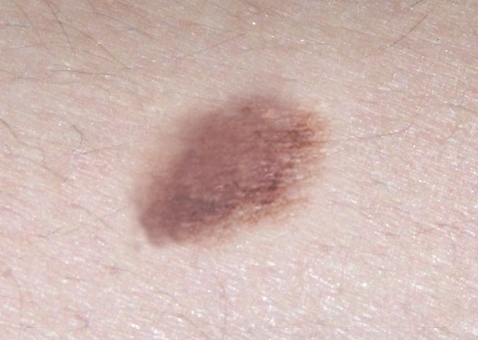
A clinically atypical mole may be larger than common moles, have irregular or blurred edges, and contain shades ranging from pink to dark brown. Sometimes a raised centre is surrounded by a flatter area. These features occur in different combinations; there is no single appearance shared by every lesion.
The diameter often exceeds 5–6 mm, but size alone cannot classify a lesion. Small moles can be atypical, and melanoma can measure less than 6 mm. The threshold is an observation aid, not a boundary between benign and malignant.
The trunk, back, abdomen and arms are possible sites. Usually covered areas, including the buttocks and breasts, can also be affected. Lack of direct sun exposure does not exclude an atypical mole. Comparing it with other moles on the same person and understanding how it has changed is more useful than comparing it only with online pictures.
What the pathologist examines
Histology examines sampled tissue rather than just visible colour or outline. Dysplastic naevi may show disordered organisation of melanocytes and atypia of individual cells, to varying degrees. Cytological atypia should therefore not be described as necessarily rare.
Increased numbers and irregular distribution of melanocytes mainly involve the epidermal–dermal junction. Uneven nests and proliferation along the basal layer may be seen. This is not simply a matter of cells “extending deep into the epidermis”. Fibrosis, lymphocytic inflammation and more prominent small vessels may occur in the papillary dermis.
The pathologist interprets the combined findings, their distribution and the context. Some lesions can be difficult to distinguish from early melanoma. Correlation with the clinical appearance, more complete sampling or a second dermatopathology opinion may then be needed. An isolated word in a report should not be treated as a stand-alone diagnosis.
Frequency and melanoma risk
Frequency estimates vary with the population studied and the criteria used. The National Cancer Institute reports that about one in ten people in the United States has at least one dysplastic naevus. This is not an Italian prevalence estimate and does not support a claim that 70% of the general population is affected.
Most dysplastic naevi do not become melanoma. Their presence, especially in greater numbers, nevertheless signals higher personal risk. These are separate concepts: the likelihood of a particular mole changing and the likelihood of a person developing melanoma, including elsewhere on the body.
Total mole count, the number of atypical lesions, personal history and family history all contribute to assessment. Published risk ratios describe groups, not an individual prediction. Calculating personal risk from a simple count or a single size threshold is inappropriate.
Melanoma can arise within an existing mole but often develops where no mole was previously visible. Removing every atypical mole therefore does not eliminate risk. Surveillance needs to cover the entire skin.
Multiple naevi, family history and genetics
One dysplastic naevus does not establish an inherited syndrome. Some people have many common and atypical moles, sometimes more than a hundred. The count alone cannot diagnose a familial syndrome. Personal melanomas, affected relatives, their relationship and age at diagnosis must also be considered.
Familial atypical multiple mole melanoma syndrome, often abbreviated FAMMM, describes particular familial patterns. In some families, susceptibility is associated with inherited pathogenic variants in CDKN2A, on chromosome 9p, or other genes. Inheritance may be autosomal dominant, but expression and penetrance vary: inheriting susceptibility does not make cancer inevitable.
Historical research into chromosomal instability, translocations and loss of chromosome 9 material contributed to understanding melanocytic biology. Changes found in cells or tissues should not be confused with an inherited variant in the person. They do not show that every dysplastic naevus follows the same sequence towards melanoma.
Genetic counselling may be appropriate for selected families, for example those with multiple melanomas or combinations of melanoma and pancreatic cancer. It is not an automatic test for every atypical mole. A specialist reviews the family history and considers the usefulness, limitations and implications of testing.
Examination, dermoscopy and monitoring
Assessment starts with the lesion’s history and a skin examination. Explain how long it has been present, how it has changed, any symptoms and previous removals. Existing photographs and pathology reports may help establish its evolution.
Dermoscopy lets the dermatologist examine structures not visible to the naked eye. In people with many moles, total-body photographs and selected dermoscopic images can support comparisons over time. Mole mapping does not replace clinical judgement or make it safe to delay assessment of a suspicious lesion.
The choice between monitoring and biopsy depends on the combined findings. A clearly suspicious lesion should not simply be scheduled for a distant check. Surveillance intervals are agreed according to risk and lesion characteristics. Ask when to return and which changes should prompt an earlier appointment.
Removal, grade of dysplasia and margins
Not every atypical mole needs removal. When a suspicious lesion requires clarification, the clinician chooses a procedure that allows adequate tissue assessment. Destructive treatments or home removal may prevent analysis and delay diagnosis.
A report may state a grade of atypia or dysplasia and whether margins are clear. “Involved margins” means lesion cells reach the edge of the sample; it does not mean metastasis. The sampling method and any remaining pigmentation also matter when deciding whether another procedure is needed.
Mildly or moderately dysplastic naevi removed with clear margins generally do not require re-excision. In selected cases with microscopically involved margins but no clinical residual lesion, a specialist may recommend observation. This option should not automatically be extended to partial biopsies, still-suspicious lesions or all grades of dysplasia.
Severe atypia or uncertainty about melanoma often leads to completion of removal or further pathology assessment. Clinician and pathologist should coordinate the decision. Keep the report and ask for an explanation of the plan: “dysplastic” alone determines neither the need for nor the extent of another operation.
Changes to report and self-examination
The ABCDE rule recalls asymmetry, borders, colour, diameter and evolution. When a person has many atypical moles, some of these features may already be present. A lesion unlike the others—the “ugly duckling”—or a change from its usual appearance is particularly useful to notice.
Report rapid growth, a new lump, a change in colour or shape, spontaneous bleeding or persistent itching. These signs do not prove melanoma on their own but warrant assessment. Do not wait for a scheduled check if a suspicious new development appears.
Skin self-examination includes the back, scalp, buttocks, feet and other areas that are difficult to see. Use a mirror or ask for help. A dated photograph may document change but does not replace an appointment. The aim is not to classify every mole yourself, but to notice changes and describe them accurately.
Skin protection and next steps
UV protection remains part of prevention: avoid sunburn and tanning beds, seek shade, wear protective clothing and apply broad-spectrum sunscreen according to its instructions. Sunscreen is not permission to prolong exposure and does not replace other protective measures.
Having a dysplastic naevus calls for attention proportionate to risk, not indiscriminate removal of all moles. After an appointment, the meaning of the report, any lesions requiring observation and the surveillance schedule should be clear. Our diagnosis guide explains the next steps, while the general page on moles provides introductory information.
Sources and update
- National Cancer Institute — Common Moles, Dysplastic Nevi, and Risk of Melanoma
- DermNet — Atypical melanocytic naevus (2025)
- National Cancer Institute — Genetics of Skin Cancer (PDQ)
- Pigmented Lesion Subcommittee — Consensus statement (2015)
- Kim et al. — Moderately dysplastic naevi with positive margins (2018)
- American Academy of Dermatology — Moles: diagnosis and treatment
- American Academy of Dermatology — When is a mole a problem?
Editorial update: 16 October 2026. This information does not replace medical assessment.