Photoageing: what Glogau describes
Skin changes with age for biological reasons, but chronic ultraviolet exposure can add to and accelerate changes in colour, texture and elasticity. This component is called photoageing. It mainly affects exposed areas such as the face, neck, upper chest and backs of the hands, with varying intensity according to exposure history and individual characteristics.
The Glogau classification is a descriptive scale of visible signs, particularly facial wrinkles. It distinguishes four grades, from early changes to very pronounced alterations. It is mainly used in the clinical and aesthetic assessment of sun damage; it does not measure the biological age of the whole body or establish whether someone has melanoma.
The ages traditionally associated with the groups are approximate. A birthday does not automatically move someone into the next grade, and a group cannot be assigned simply from a photograph. Assessment considers the overall findings and examination of the skin.
The four grades of photoageing
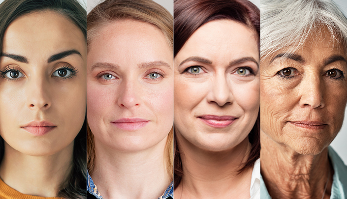
Group I — early changes. Permanent wrinkles are absent or minimal. Mild pigmentation irregularities may appear, while surface changes are subtle. The classical description does not include obvious keratoses. This category is often illustrated using young adults, but age is not the deciding criterion: skin without wrinkles is not necessarily skin that has never sustained sun damage.
Group II — wrinkles mainly with movement. Photoageing becomes more noticeable during smiling and other expressions. Solar lentigines, early texture changes and more visible lines in mobile facial areas may appear. Traditional descriptions note that some keratoses may be felt before they are clearly visible. A persistent rough area, however, should be assessed as a lesion in its own right, rather than simply classified as an aesthetic change.
Group III — wrinkles also at rest. Lines remain visible when the face is not moving. More marked uneven pigmentation, dilated superficial vessels and surface changes, including visible keratoses, may coexist. Damage is more advanced, but the number of wrinkles cannot establish the nature of a spot or nodule in the same area.
Group IV — very pronounced changes. Wrinkles are widespread and deep, with few apparently smooth areas; yellowish or greyish colour and substantial texture irregularity may coexist. Classical descriptions sometimes include a history of skin cancers or precancerous lesions. This does not mean everyone in the group has cancer, or that cancer only appears at this stage. Difficulty achieving an even make-up application is an aesthetic observation, not an oncological diagnostic criterion.
Glogau, Fitzpatrick and melanoma risk: three different questions
Glogau mainly describes what is visible on the skin after exposure and ageing. The Fitzpatrick classification instead describes the tendency to burn and tan. Melanoma risk factors include additional elements such as mole count, family history, previous melanomas and immunosuppression. The three assessments are not interchangeable.
A person with few wrinkles may have a history of sunburn or significant inherited susceptibility. Conversely, severely photoaged skin does not allow a melanoma diagnosis without examination of individual lesions. The scale justifies neither automatic reassurance nor alarm based solely on facial appearance.
This distinction also helps interpret images: the faces shown together in the illustration are not diagnoses, predictions of disease or an inevitable sequence for everyone. Sun protection and checks should be chosen according to the clinical situation, not an aesthetic comparison with a photograph.
Spots and keratoses: when examination is needed
A brown spot may be a solar lentigo, but it needs to be distinguished from other pigmented lesions. A rough area may be an actinic keratosis, which mainly belongs to the squamous cell carcinoma pathway and should not be described as early melanoma. Not all “age spots” are alike, and treating them as one cosmetic problem is unwise.
A new or changing lesion, a growing nodule, unexplained bleeding or a small wound that fails to heal needs dermatological assessment. The ABCDE rule can help identify suspicious changes in pigmented lesions, but it does not cover every presentation of skin cancer.
Before destructive cosmetic treatment of a spot or uncertain lesion, its nature should be established. Dermoscopy and, when indicated, histological examination may be needed. Lightening a spot without a diagnosis is not the same as appropriately treating a possible disease.
Reducing future damage and planning checks
Accumulated damage does not make prevention pointless. Protecting the skin from UV radiation helps reduce further exposure: shade, clothing, a hat and broad-spectrum sunscreen on uncovered areas should be considered together. General recommendations include SPF at least 30 and regular reapplication; no product permits longer sun exposure, and tanning beds are not a safe alternative.
The frequency of examinations depends on previous skin cancers, moles, family history, immunosuppression and clinical findings. Between visits, skin self-examination helps identify new changes, including in hard-to-see areas. A changing lesion should not wait for a cosmetic appointment or the next already scheduled check.
Photoageing treatments may have aesthetic or functional goals, but they require individual selection and do not replace cancer surveillance. This page explains a classification: it does not recommend a treatment based solely on a Glogau group.
Sources and update
- Glogau RG — Aesthetic and anatomic analysis of the aging skin (1996)
- Richard Glogau — Glogau Wrinkle Scale
- WHO — Ultraviolet radiation
- American Academy of Dermatology — Sunscreen FAQs
- NCI — Skin Cancer Prevention (PDQ)
Editorial revision: 17 October 2026. This material is educational and does not replace medical assessment.