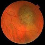
Choroidal melanoma: it is a tumor that originates from the melanocytes of the choroid below the retina and represents the most frequent malignant tumor of the eye, it has an incidence of about 1.5% out of 100,000 people per year (400 new cases in Italy every year) with a higher frequency in males between 50 and 60 years, rare among adolescents and Hispanic races.

In most cases it arises ex novo, in rare cases it arises from a nevus. The 5-year mortality rate after enucleation of the eye is approximately 60% with a maximum mortality 2 years after enucleation. In the case of metastases, survival is comparable to that of cutaneous melanoma. In the 1990s some chromosomal alterations in uveal melanoma were highlighted, in particular polysomy of chromosome 8 and monosomy of chromosome 3. The identification of alterations can occur by FISH (fluorescence in situ hybridization) and is associated with an unfavorable prognosis. The tumor is often asymptomatic in the initial stages and is identified during a routine examination for which it is advisable for everyone to perform a periodic visit including ophthalmoscopy with dilation; when symptomatic, flashes or floating vision (retinal detachment), distortions of vision, loss of vision (involvement of the fovea or parafoveal area) appear. If it affects the anterior region of the eye near the lens, it can give rise to irregular astigmatism. In the case of iridociliary or anterior choroidal melanoma, brownish patches visible to the naked eye, irregularly shaped pupil and secondary glaucoma may be noted. Normally melanoma is pigmented, however there are cases of choroidal amelanotic melanoma, the orange pigmentation with lipofuscin is a sign of metabolic activity.
The diagnosis occurs mainly with the ophthalmoscope (indirect binocular ophthalmoscopy) together with the clinical examination and ocular ultrasound (A-B scan technique) which allows to evaluate the thickness of the tumor. In doubtful cases, CT, MRI and finally fine needle biopsy are used. In the suspicion of a metastatic dissemination it is necessary to perform a total body CT or PET and a study of the liver parameters given the frequent localization in this site. The differential diagnosis is with choroidal hemangioma, choroidal nevus, RPE hypertrophy, subretinal hemorrhages, choroidal metastases, melanocytoma, and choroidal osteoma. The prognosis varies according to the histological subtype with a greater aggressiveness for the spindle cell form, furthermore the mitotic index and the possible invasion of the sclera or anterior chamber, necrosis, lymphocytic infiltration and the prominence of the nucleoli. Based on the thickness and diameter of the base, they are divided into small (S=1-2.5 mm; L=5 mm), medium (S= 2.5-10 mm; L= 5-16 mm), large (S=>10 mm; L= >16 mm).
Histologically, 3 variants are distinguished: spindle cell (2 subgroups (A with elongated nuclei with nucleolus and longitudinal notch from invagination of the nuclear membrane and type B with rounded nuclei and evident eosinophilic nucleolus), epithelioid cells (polygonal cells with large nucleus and with obvious eosinophilic nucleolus and abundant cytoplasm) and a mixed cell type. From a therapeutic point of view, we recommend enucleation of the eye, radiotherapy and in selected cases photocoagulation and transpupillary thermotherapy (increase of temperature inside the tumor with a diode laser).In medium-sized tumors, brachytherapy with iodine 125 or ruthenium 106 episcleral plates has been proposed with results similar to enucleation of the globe and proton therapy with photons or helium. Globe enucleation is advisable, radiation therapy is sometimes done, but vision is often impaired and subsequent pain often leads to subsequent enucleation surgery.
The most frequent metastases are to the liver and sometimes also to the kidney, ovary, lung and other organs with little involvement of the lymph node districts. In the case of metastasis, the therapy is not dissimilar to that of cutaneous metastatic melanoma with chemotherapy, vaccines and immunotherapy. Photoemustine appears to be more effective in metastatic choroidal melanoma and treatments with intra-arterial hepatic perfusion have been proposed.
With regard to prevention, the use of glasses with protective lenses against UV rays is recommended as a greater incidence has been seen among patients with blue or light eyes, who carry out outdoor activities, for which it is believed that excessive exposure to UV radiation favors the development of choroidal melanoma.