What acral lentiginous melanoma is
Acral lentiginous melanoma arises from melanocytes at acral sites: the palms, soles and nail unit. “Lentiginous” describes a pattern of cell growth, not an ordinary freckle.
It is relatively rare in fair-skinned populations but represents a larger proportion of melanomas diagnosed in dark-skinned and some Asian populations. This compares the distribution of subtypes; it does not mean melanoma overall is more common in people with dark skin.
It affects both sexes and different ages, particularly adults and older people. Widely varying study percentages should not be applied directly to an individual or used to exclude suspicion in other groups.
Location and relationship with sunlight
The soles and other less visible areas can delay recognition. Nail-unit disease is also covered in the nail melanoma page because it presents particular diagnostic and surgical issues.
Unlike many melanomas on sun-exposed skin, acral melanoma has no established relationship with UV exposure. Its causes are not fully understood and should not automatically be attributed to injury, footwear or friction. Sun protection remains important for skin health but does not replace observation of palms, soles and nails.
Possible appearances
One possible presentation is an enlarging brown or black patch with irregular shape, edges or colour. A raised or nodular component may subsequently develop. Poorly pigmented, pink or ulcerated lesions resembling a nonhealing wound also occur.
Calluses, warts, bruises and other benign conditions can complicate recognition. A persistent change or a lesion not responding as expected to treatment warrants reassessment. Pain does not have to be present.
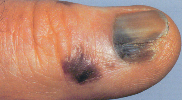
The archive photograph shows a digit with nail-area involvement; it does not represent every acral presentation. No photograph alone reliably distinguishes melanoma from a benign lesion.
Dermatological assessment and biopsy
Examination considers location, evolution, pigmentation and the person’s other lesions. Dermoscopy examines pigment distribution in relation to acral skin structures; interpreting these details requires specific experience.
Histological examination confirms the diagnosis. Biopsy technique depends on extent and location, aiming for adequate sampling even in functionally delicate areas. A superficial or unrepresentative sample may not describe the whole lesion.
The report establishes invasion, Breslow thickness, ulceration and margins. These findings inform consideration of sentinel lymph node biopsy and other investigations. Clinical inspection alone cannot determine stage.
Treatment and hand or foot function
Surgery is central to localised disease. Planning must obtain adequate margins while considering weight-bearing, mobility, healing and reconstruction. Acral location does not automatically mean amputation; decisions depend on extent and the structures involved.
Higher-risk or advanced cases may be considered for immunotherapy and targeted treatment when actionable molecular alterations are present. Radiotherapy and chemotherapy have selected roles. Biology may differ from other melanomas, so treatment is based on the findings in the individual case.
Prognosis depends mainly on stage and pathological features. Delayed recognition can contribute to more advanced diagnosis but does not determine every patient’s outcome. During skin self-examination, include the soles, spaces between digits and nails, and report new changes promptly.
Sources and update
- National Cancer Institute — Melanoma Treatment (PDQ)
- ESMO — Cutaneous melanoma: Clinical Practice Guideline (2025)
- DermNet — Acral lentiginous melanoma
Editorial revision: 16 October 2026. This information does not replace medical assessment.