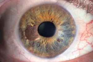
Differentiating a melanoma from an iris nevus is extremely difficult and sometimes impossible. Melanoma is often a small lesion and rarely appears infiltrative, multiple, and can cause chronic uveitis and heterochromia.

If they affect at least 66% of the circumference angle, melanoma can cause glaucoma. The investigations to be performed are transillumination, indirect ophthalmoscopy, gonioscopy. Photographic documentation allows you to follow the progression of the tumor. Fluorescence angiography of the anterior segment evaluates vascularity but is not diagnostic.
High-resolution ultrasound biomicroscopy is used to measure the lesion and to evaluate the involvement of the ciliary body, angle, or sclera. The median survival is about 95% at 5 years. Treatment is conservative when possible. In asymptomatic patients, photographic documentation is recommended if the lesions are substantially stable. If the tumor is growing, resection is recommended.
Enucleation is recommended in cases where local surgical treatment is not feasible.
Plaque radiation therapy is used in inoperable cases.