Rhabdoid does not mean muscle origin
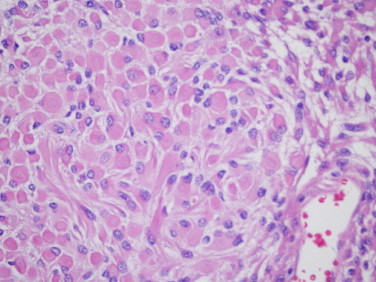
Rhabdoid melanoma contains cells resembling those in other rhabdoid neoplasms: abundant cytoplasm, eosinophilic inclusions and a nucleus often displaced towards the edge. The term is morphological. It does not prove muscle origin and is not equivalent to rhabdomyosarcoma.
This appearance is described particularly in recurrent or metastatic settings, although primary tumours have been reported. It can coexist with other melanoma components in the same sample. Biopsy location and cancer history are therefore essential.
Microscopic features
Cells may be polygonal and arranged in sheets, with vesicular nuclei and conspicuous cytoplasmic inclusions. Melanin may be sparse or absent, making melanocytic origin less obvious. Lack of pigment cannot exclude melanoma.
Histological examination also considers areas without rhabdoid features. A limited sample may show only the most unusual component, complicating distinction from carcinomas, sarcomas or other poorly differentiated neoplasms. A microscopic photograph illustrates morphology but cannot replace assessment of the complete specimen.
Interpreting markers in context
Expression of S100 and vimentin has been described, sometimes with unexpected markers such as keratins or desmin. These findings are not an invariant signature. Vimentin is nonspecific, and a marker usually associated with another lineage cannot resolve the diagnosis alone.
The panel may include SOX10 and other melanocytic markers alongside those needed to exclude alternatives. Dedifferentiated melanomas can lose some markers. Comparison with a previous melanoma, expert review and selected molecular investigations help integrate the findings.
The report should explain diagnostic certainty and distinguish the rhabdoid component from disease stage. These are different pieces of information.
Treatment and prognosis without overgeneralisation
Many published cases concern aggressive or already advanced disease. This does not prove that rhabdoid appearance always causes poor response to every treatment independently of stage. Rarity limits reliable comparisons and individual predictions.
Treatment follows the melanoma diagnosis, site and extent. Surgery, immunotherapy, appropriate targeted treatments and radiotherapy have different roles according to the case. Chemotherapy may be considered in selected settings, not automatically because of morphology.
An expert centre can discuss possible clinical trials and plan surveillance and support. Useful questions concern lesion origin, spread, molecular results and the concrete goal of treatment.
Sources and update
Editorial revision: 17 October 2026. This information does not replace medical assessment. For very rare variants, some evidence comes from small series or case reports.