Educational information, not a prescription. Decisions require specialist assessment; editorial source checking is not equivalent to clinical validation.
How to read this atlas
The atlas retains photographs from the original section curated by Dr Daniele Gandini. They document individual cases and different moments in care; they cannot establish a personal diagnosis, select a technique or predict an outcome. Captions have been editorially revised to prevent historical descriptions becoming universal recommendations.
Photographs include lesions, wounds and operations that some readers may find distressing. Each group relates the archive description to current context. Where complete clinical information is unavailable, no stage, long-term outcome or definite cause of a complication is inferred. General clinical context is linked to the cited sources and the page on plastic and reconstructive surgery for melanoma.
Sources: ESMO — Cutaneous melanoma: Clinical Practice Guideline (2024/2025); Cancer Research UK — Surgery to remove melanoma skin cancer.
Excision: from historical operations to current planning
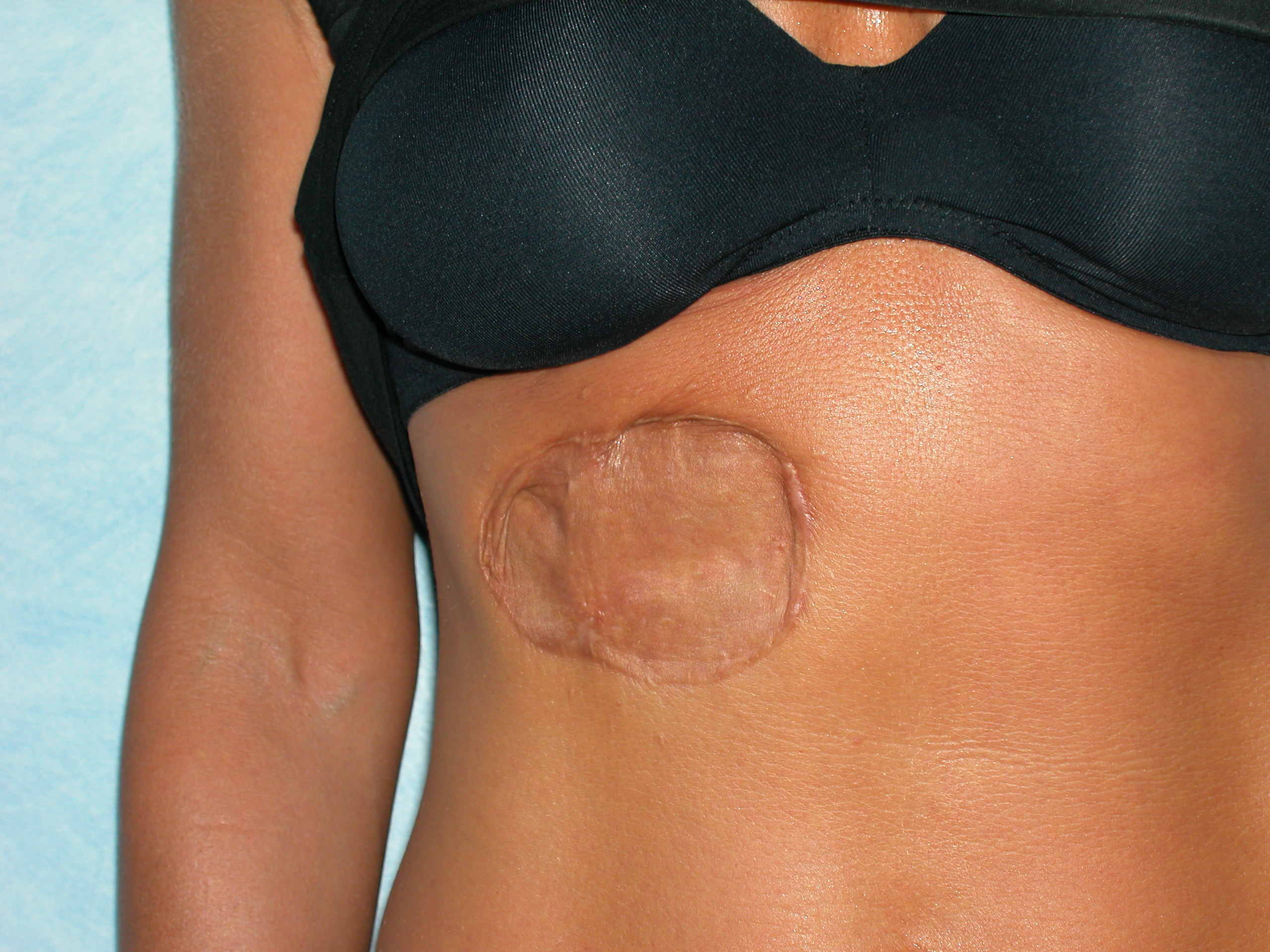
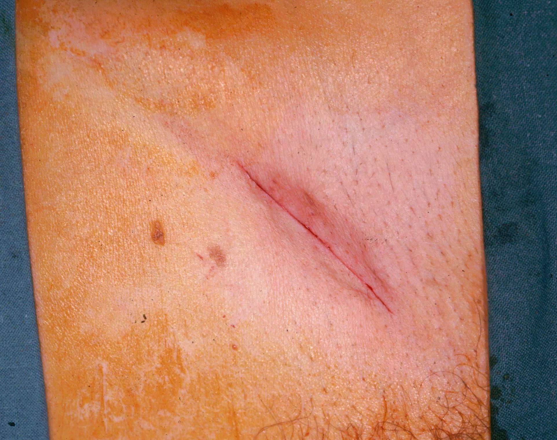
The first two images illustrate outcomes after different excisions. The first was described in the archive as the result of a wide Olsen-type excision; the second shows a linear scar. They are not a controlled comparison and do not show that the second result is achievable in every situation.
Today, excision extent is based on histological characteristics and guidelines, with attention to site and function. The priority is adequate cancer treatment while limiting unnecessary tissue loss. Highly extensive historical operations should not be presented as the routine model, but neither should they be replaced by a promise of invariably minimal scars.
Sources: ESMO — Cutaneous melanoma: Clinical Practice Guideline (2024/2025).
Local flap: planning and immediate appearance
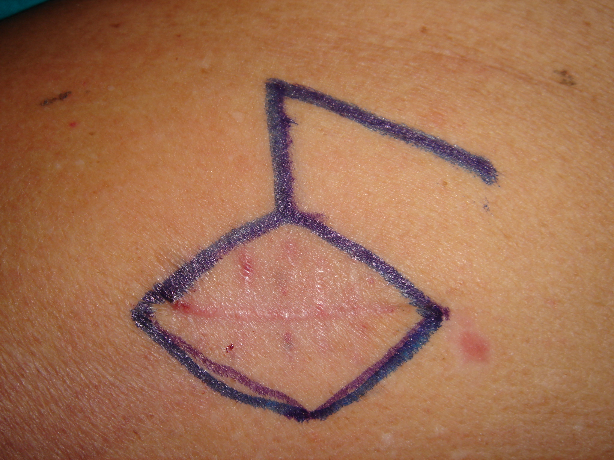
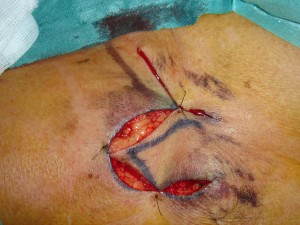
This sequence retains the design of a Limberg flap, tissue transfer and suturing. A flap uses tissue with a blood supply to cover a defect. Its design must respect anatomy, tension and neighbouring structures; reproducing the drawing on skin is not sufficient.
The final photograph shows the immediate postoperative appearance, not the definitive scar. Healing, possible complications and scar maturation can change appearance over time. These images cannot guarantee absence of distortion, tension or a need for later revision.
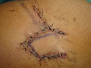
Sources: Cancer Research UK — Surgery to remove melanoma skin cancer; Cambridge University Hospitals — Care of your scar.
Grafts and donor areas
A graft may be an option when direct closure is unsuitable. This group combines an outcome illustrated in the archive with two examples of donor sites. Interpretation requires distinguishing full-thickness and split-thickness grafts: tissue characteristics and donor-site management differ.
The groin is one possible source for small full-thickness grafts, not the ideal choice for every area. Thinner grafts can cover extensive surfaces but are not reserved exclusively for trauma. Selection depends on the recipient bed, dimensions, function, required colour and thickness and the patient’s condition. The donor site also leaves an outcome that should be discussed.
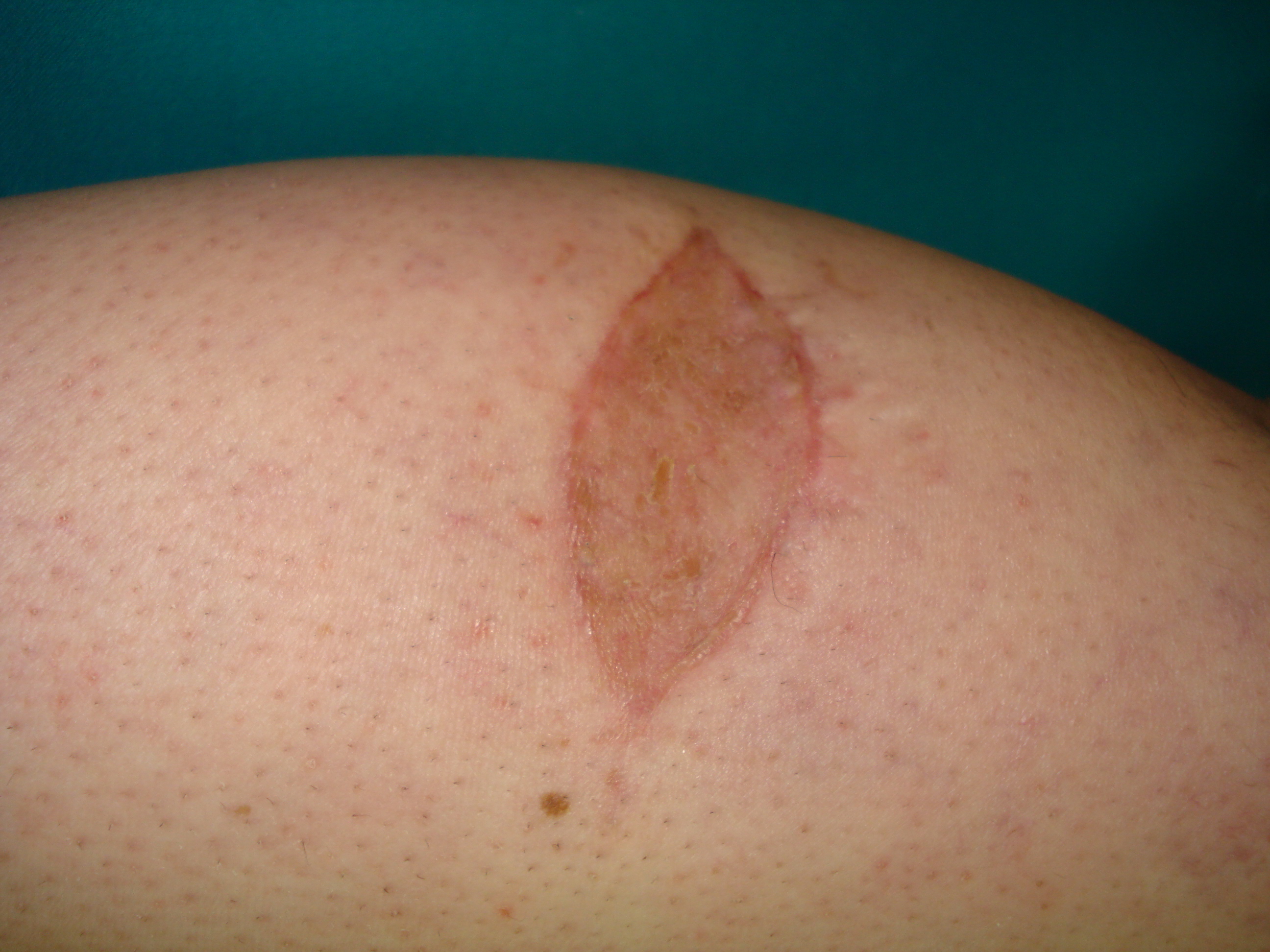

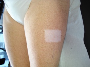
Sources: Cancer Research UK — Surgery to remove melanoma skin cancer.
Sentinel lymph node localisation
These images show dye use, an operative field and equipment for intraoperative detection. Radiotracer and probe help identify first-draining lymph nodes; dye may provide additional guidance according to the protocol.
Sentinel lymph node biopsy provides staging information when indicated. It is not the same as lymph-node clearance and does not eliminate false-negative results or complications. A device photograph does not certify procedural accuracy. Indication, coordination with excision and interpretation of results belong to the specialist pathway described in the dedicated guide.
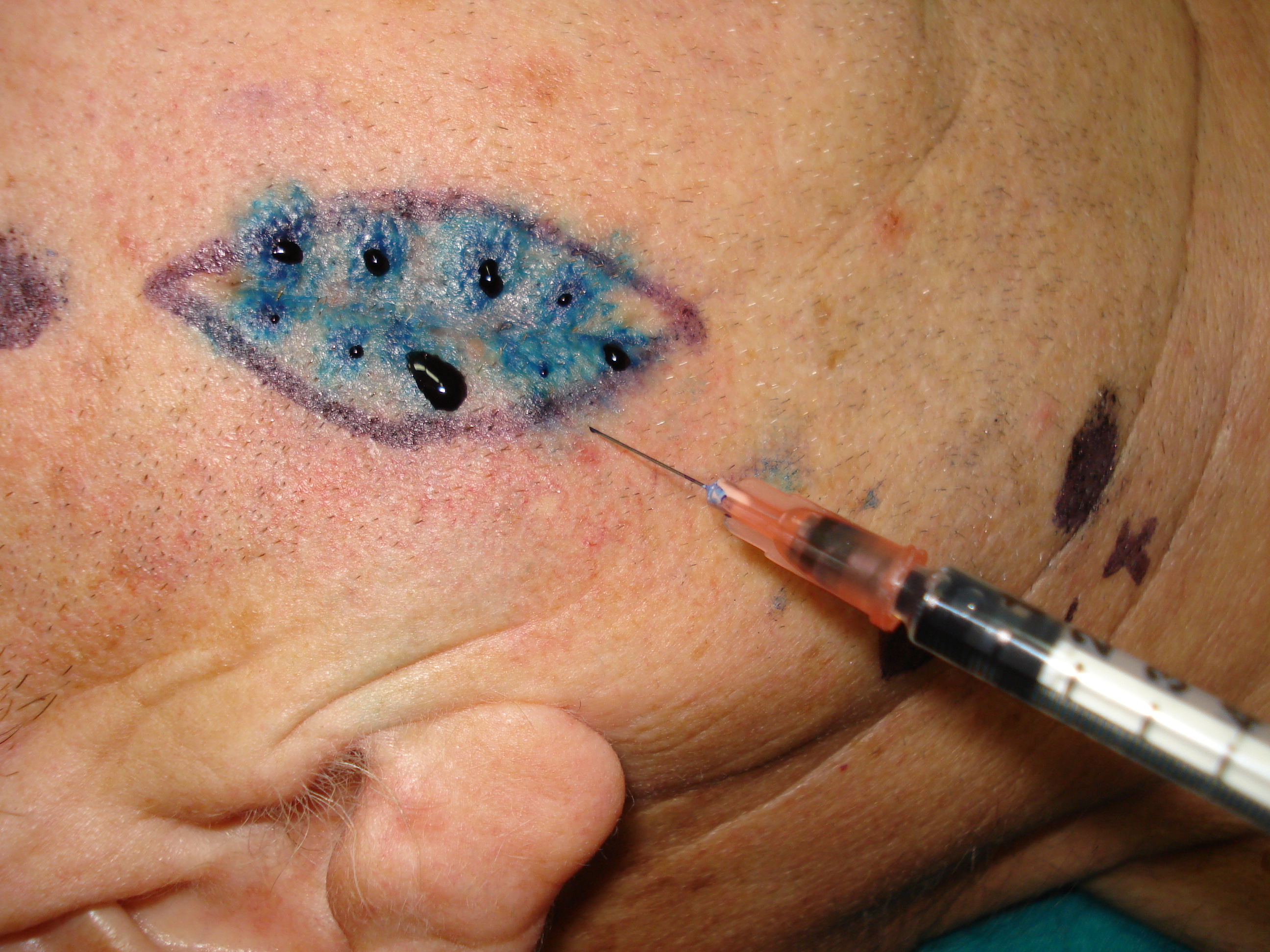
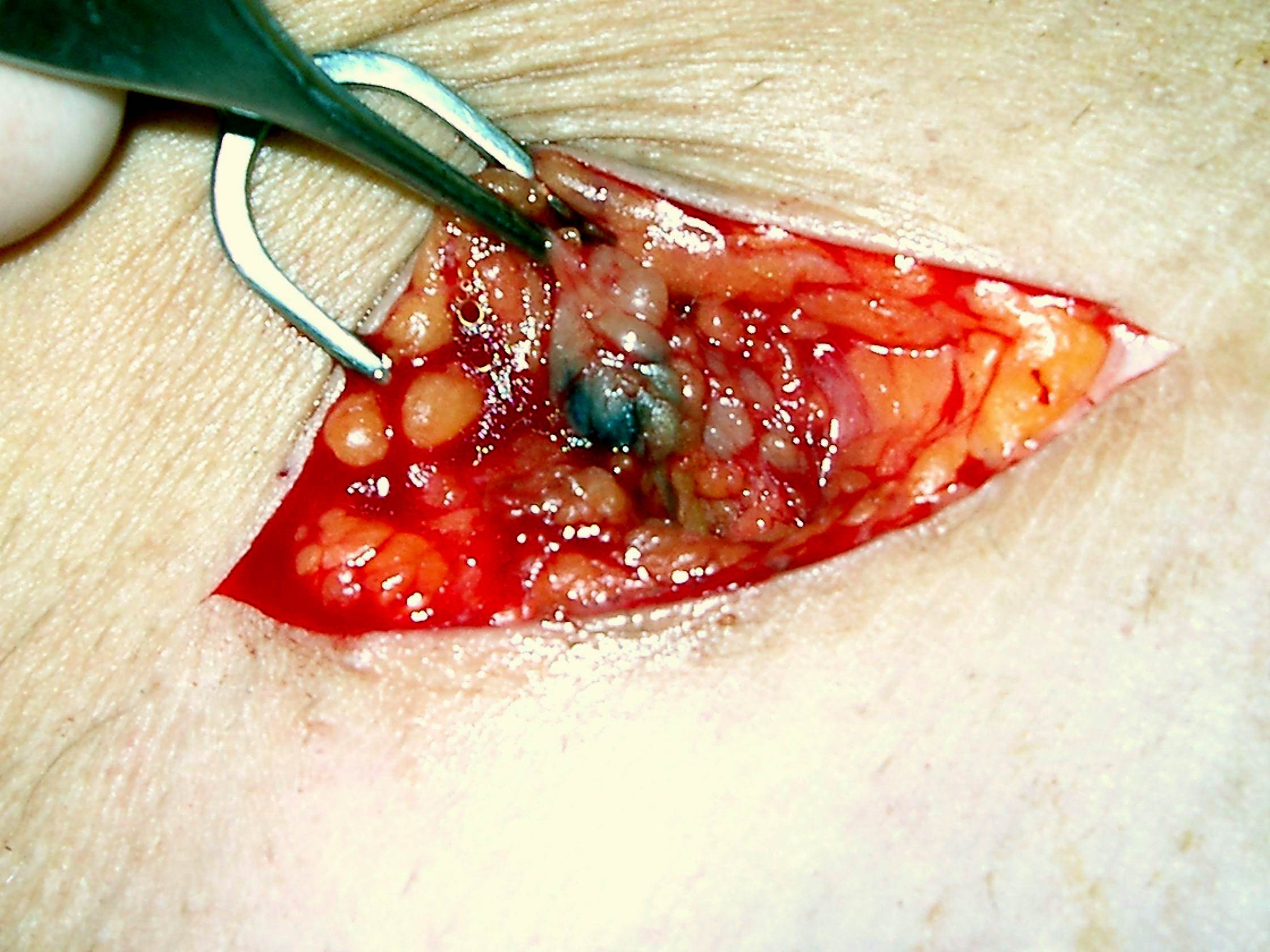
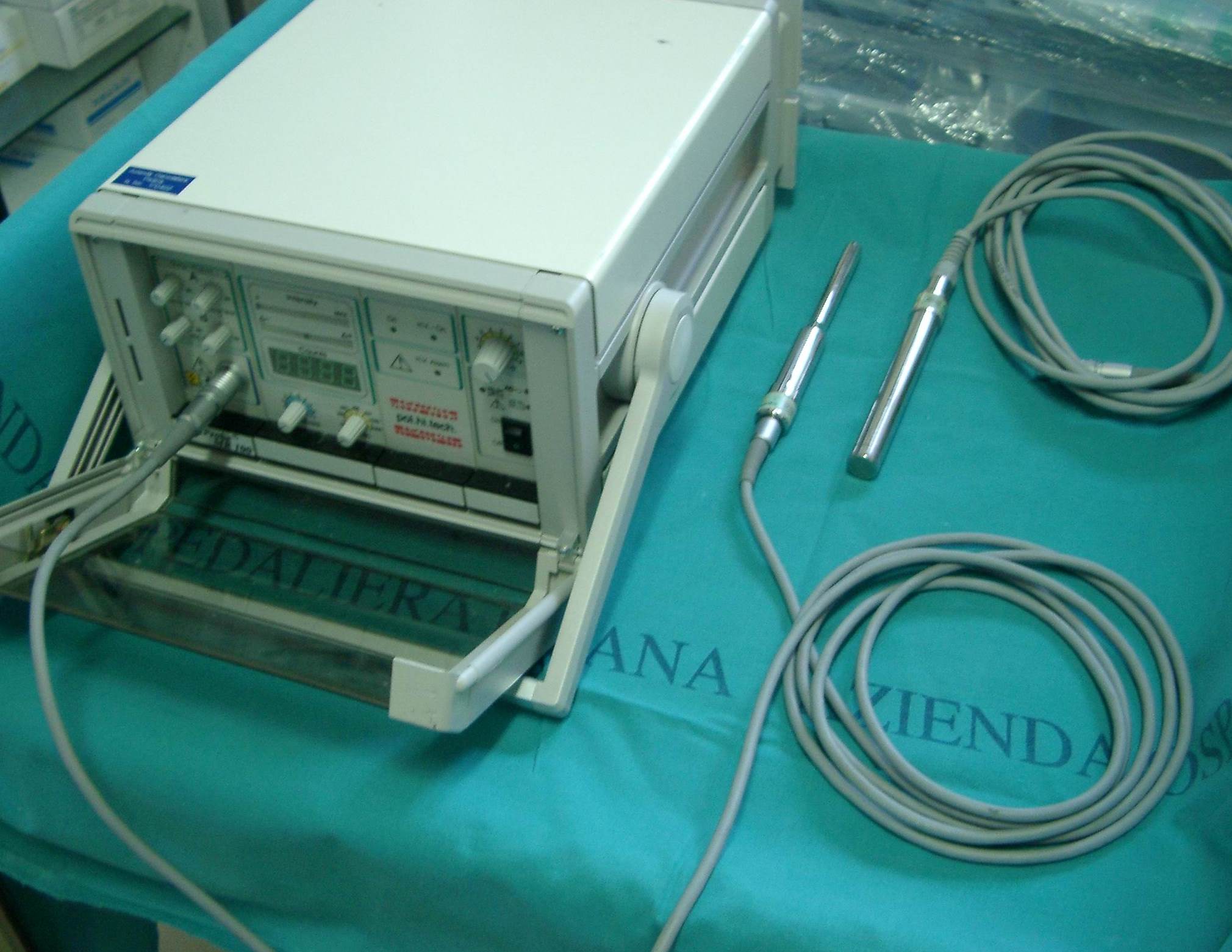
Sources: ESMO — Cutaneous melanoma: Clinical Practice Guideline (2024/2025); NICE NG14 — Melanoma: assessment and management.
Melanoma with clinically apparent nodal enlargement
The archive described this case as nodular melanoma of the arm with axillary lymph-node enlargement. Suspicious nodal swelling requires diagnostic confirmation and staging: this is not a routine situation addressed solely by sentinel-node localisation.
The historical caption called for immediate nodal clearance. Today, sequencing requires multidisciplinary discussion: neoadjuvant treatment may be appropriate in some resectable stage III cases according to evidence and access criteria. The photograph cannot establish this individual case’s eligibility for present-day options.
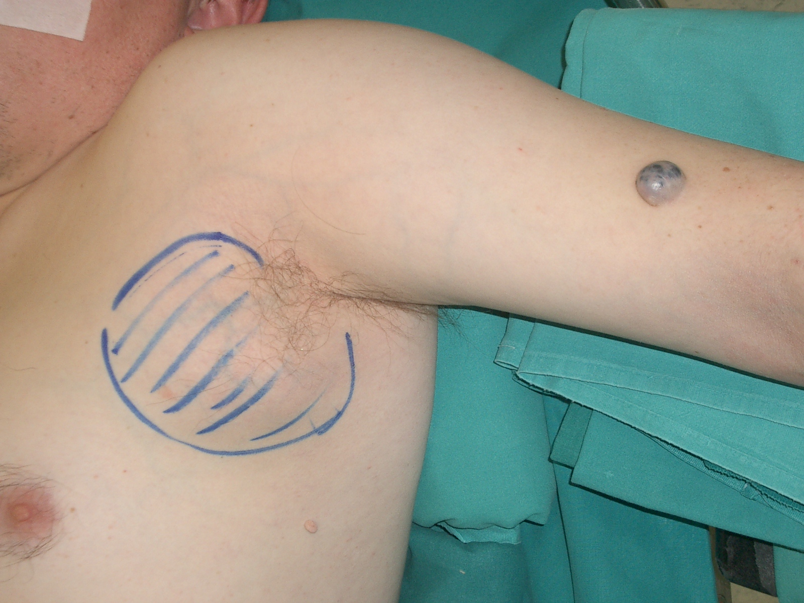
Sources: NADINA — Neoadjuvant nivolumab and ipilimumab in resectable stage III melanoma (2024); AIFA — Neoadjuvant nivolumab–ipilimumab, Law 648/1996 (17 November 2025); NICE NG14 — Melanoma: assessment and management.
Extensive foot lesions and preserving function
The photograph documents a large pigmented forefoot lesion for which the archive described a major ablative operation. It cannot independently establish diagnosis, depth or the need for amputation. Before irreversible surgery, clinical assessment, histological confirmation and oncological and reconstructive planning are required.
At acral and nail sites, balancing margins, coverage and function requires particular expertise. A targeted partial biopsy may be selected when complete diagnostic removal would be disproportionate. Tissue-preserving options are not always feasible, but a historical case should not become a universal indication for amputation.
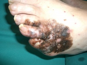
Sources: ESMO — Cutaneous melanoma: Clinical Practice Guideline (2024/2025); JCOG1602/J-NAIL — Protocol for non-amputative digit-preserving surgery (2019).
Recurrence and symptom control
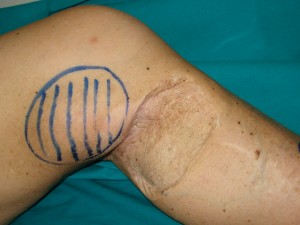
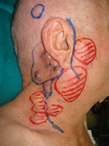
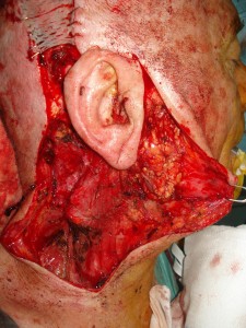
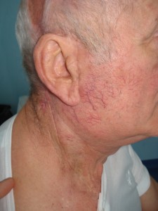
The first image was described as a subcutaneous recurrence near the knee. The following three belong to archive sequence 12a–12c, concerning surgery for advanced disease in the head-and-neck region. They are kept together to preserve the documentation’s context without assigning an unavailable oncological outcome.
Surgery for recurrence or metastases can have different goals: removal of selected disease, local control or reduction of bleeding, ulceration or pain. A lesion appearing solitary in a photograph does not establish that it is the only disease site. Staging and its relationship with immunotherapy, targeted treatments or other approaches are essential to decision-making.
Sources: NICE NG14 — Melanoma: assessment and management; Cancer Research UK — Surgery to remove melanoma skin cancer.
Complications and new lesions near a scar
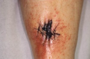
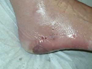
The leg photograph was described as skin necrosis; the next as satellite lesions near the operated site. These are different problems: the first concerns tissue viability and healing, while the second requires cancer assessment. An image cannot attribute a complication to surgical error or confirm the nature of a new lesion without investigation.
Satellite nodules or lesions are not necessarily dark: they may be small or reddish. A change near the scar warrants assessment and should not automatically be considered a normal consequence of surgery. Increasing pain, fever, discharge, bleeding or important wound changes should be reported to the centre. Clinical follow-up complements this photographic documentation rather than being replaced by it.
Sources: Cancer Research UK — Surgery to remove melanoma skin cancer; NICE NG14 — Melanoma: assessment and management.
Sources and update
- ESMO — Cutaneous melanoma: Clinical Practice Guideline (2024/2025)
- Cancer Research UK — Surgery to remove melanoma skin cancer
- Cambridge University Hospitals — Care of your scar
- NICE NG14 — Melanoma: assessment and management
- NADINA — Neoadjuvant nivolumab and ipilimumab in resectable stage III melanoma (2024)
- AIFA — Neoadjuvant nivolumab–ipilimumab, Law 648/1996 (17 November 2025)
- JCOG1602/J-NAIL — Protocol for non-amputative digit-preserving surgery (2019)
Editorial revision and source consultation: 17 October 2026. The original contribution is distinguished from the editorial update; no renewed specialist clinical review is certified.