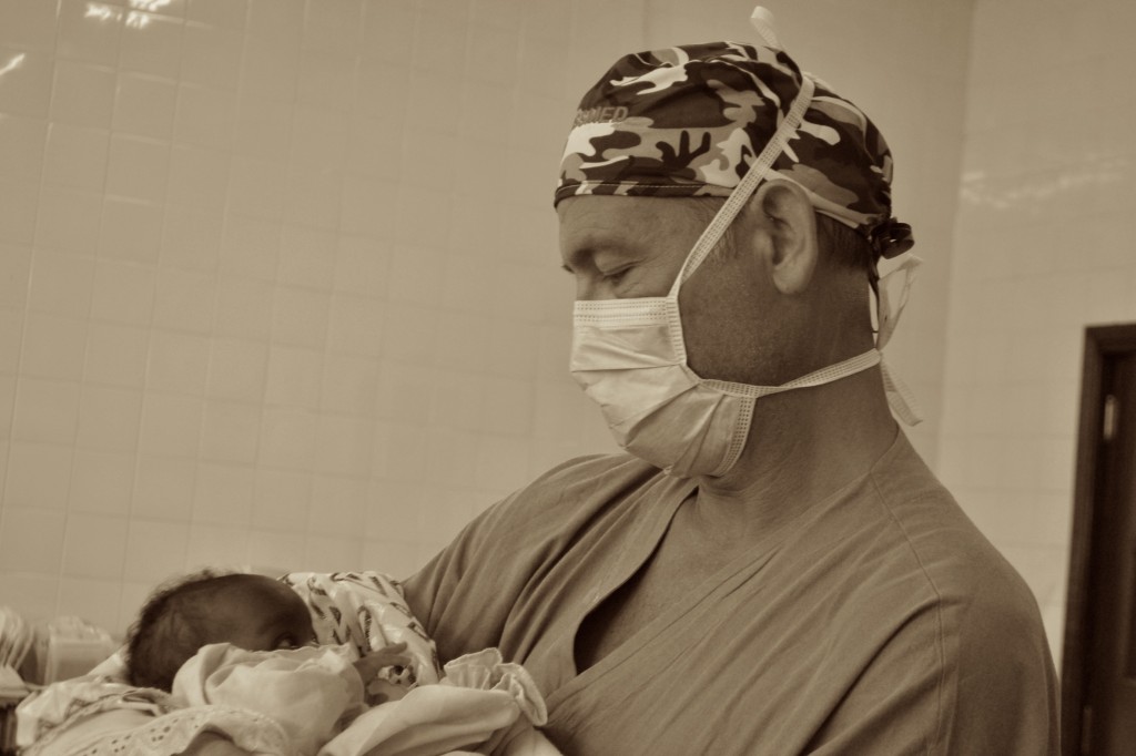
Melanoma plastic surgery is a highly specialized medical field and crucial in the management of this dangerous form of skin cancer. Melanoma is one of the most aggressive skin cancers, but when diagnosed at an early stage, treatment options, including plastic surgery, can offer remarkable aesthetic and functional results. Plastic surgery in the treatment of melanoma aims to remove the tumor while preserving the best possible functionality and aesthetics of the affected area. Thanks to the continuous advances in surgical technology and reconstruction techniques, plastic surgery specialists are able to provide customized solutions for each patient, improving the quality of life and contributing to the success of the treatment. This introductory paragraph explores the importance of plastic surgery in the management of melanoma, emphasizing the vital role it plays in ensuring both patient survival and complete aesthetic and functional recovery.
Section edited by Dr Daniele Gandini, Specialist in Plastic, Reconstructive and Aesthetic Surgery, S. Rossore Nursing Home, Pisa
(booking info: 050586217 o 050586111 – mobile: 3386053169)
Excisional biopsy: The first surgical approach to a suspected pigmented skin lesion must always be that of a limited excision for biopsy purposes (1-2 mm margin) and direct suture. As we will see later, a directly extensive removal, as was in use years ago, is absolutely to be avoided, given that the wider scar result, would irreparably compromise any (where indicated) sentinel lymph node search procedure (see section “Treatment melanoma surgery”); this is because a scar of a certain size damages the anatomy of the regional lymphatic drainage, the integrity of which is necessary for the correct migration of the radiopharmaceutical used by the surgeon to identify the lymph node (or lymph nodes) draining the melanoma area. Even the small scar from the first excision for bioptic purposes must in any case always be done in absolute compliance with plastic surgery techniques, i.e. with extreme delicacy in the manipulation of the tissues, possibly without any lateral dissection, and with rapid and very accurate hemostasis, to avoid risks of spread of any malignant cells by blood. The excision of a suspected melanoma must always be complete and full thickness except in very rare selected cases, for example of large lesions (extensive lentigo malignant, Dubreuilh ..), where to avoid disabling demolitions in the absence of a certain diagnosis, it can be an intralesional biopsy is required, a procedure that may carry some risk of metastatic spread. The limited excision of the suspected nevus must always be performed with a cold scalpel (not electrosurgical or laser!) and can generally be done on an outpatient basis, under local anesthesia, with anesthetic infiltration, possibly with the addition of a vasoconstrictor (adrenaline) without infiltrating the area below the lesion but only at the margins (so-called barrier anesthesia). The closure of the small cutaneous lozenge requires, practically in any cutaneous district, only a direct suture both of the deep planes (vycril, dexon, maxon, biosyn, nylon) and of the skin (nylon). Unless strictly necessary (large lesions in particular areas), local flaps and even less skin grafts in the first instance are absolutely to be avoided, again due to the high risk of compromising the lymphatic pathways.
Enlarged excision: In the event of histological confirmation of melanoma, an enlargement of the limited excision width according to current guidelines should be provided as soon as possible, and in any case no later than three months. In this case, depending on the area, it may be necessary to perform proximity flaps (Limberg, Doufourmentel) or full-thickness skin grafts (according to Wolfe), permitted in this phase, given that any marking of the lymph node with contrast medium has already been done. The surgical radicalization (enlargement) procedure is in fact performed immediately after lymphoscintigraphy (same day or day after), at the same time as selective lymphadenectomy (removal of the sentinel lymph node). In any case, the simplest possible procedures must always be adopted to repair the loss of residual substance and direct closure, where possible, is always preferable. The use of distant island flaps is exceptionally to be reserved only for cases of exposure of bone, vascular or tendon structures. Even if aesthetically not valid, the graft is preferable to local flaps, since it can facilitate the identification of any satellite metastases in case of locoregional recurrences. The current enlargement margins are decidedly reduced compared to the past, just think that only 15 years ago the so-called enlargement was still being performed with the technique of Olsen (Scandinavian surgeon of the seventies) which provided for extensive excisions of 10 cm and more, even if with the sparing of the muscle fascia. The current reductions (maximum 3 cm) allow in the vast majority of cases to perform an adequate closure of the area, but some sensitive areas, such as for example the face and ear, may require small changes to the protocols, with a reduction in the safety margins, in order to allow an adequate reconstruction of the area, without causing psychologically disabling mutilations; here too, plastic surgery has many reconstructive solutions, such as for example in the ear, where particular techniques allow remodeling without distortions even after an enlarged excision of melanoma or in the nose where it is possible to preserve its aesthetic unity. In the case of subungual melanoma of the hand and foot, the guidelines impose the disarticulation of the entire distal phalanx; for proximal finger melanomas, complete finger amputation is indicated if adequate excision margins cannot be performed involved including the metacarpal for the hand and the metatarsal for the foot.
Sentinel lymph node biopsy: This procedure, whose characteristics and indications are already extensively described in the “surgical treatment” section, requires some important precautions on the part of the plastic surgeon who performs it. Depending on the number of lymph nodes identified and the locations, a short general anesthesia may be required, but it is usually done under sedation plus local anesthesia. The lymph node station is infiltrated with an anesthetic solution (bupivacaine, mepivacaine,…) with a vasoconstrictor, diluted with physiological solution. The radiopharmaceutical used will allow an expert operator, assisted in the operating room by the nuclear doctor, and with the use of an intraoperative scintigram detector, to remove the lymph node without problems and with very high precision. The additional use of a vital dye (patent blue V, more suitable than methylene blue) can further “visually” facilitate the surgeon’s task in locating the lymph node. The vital dye must be injected in the same places as the radiopharmaceutical (around the scar) about twenty minutes before surgery. The sentinel lymph node procedure must also be performed in full compliance with the basic rules of plastic surgery, i.e. maximum atraumaticity for the tissues, minimum bleeding and respect for the healthy structures surrounding the lymph node to be removed; a delicate and well-performed operation reduces any possible local complications of this procedure to almost zero: haematomas, seromas, lymphedema of the limb). At the end of the operation, a small laminar drainage (Penn Rose) must always be positioned for a few days to avoid lymphatic collections. These are interventions that can generally be done in a day hospital regime, even in the case of general anesthesia. Adequate perioperative antibiotic prophylaxis is always recommended. Contraindications to the execution of this procedure are finally, in addition to previous non-limited removal with the use of flaps or grafts, the presence of suspected lymph node adenopathies, evidence of distant and/or visceral metastases, serious associated pathologies. A relative contraindication is obesity.
Scarring: With current excision margins being minimal compared to past years, old and unsightly scarring after melanoma surgery is minimized, but in some cases, patients (especially young women) may be required to correction of residual scars. For obvious oncological reasons (monitoring of possible satellite recurrences) it is not recommended to carry out corrective procedures especially if with tissue injection (flaps, lipofilling) within the obligatory 10 years of follow up (monitors over time); some cases of subtle, i.e. low-risk melanomas, may require aesthetic-reconstructive procedures even after five years, but never before.
Bibliography
1 Borgognoni L , Clinical recommendations for cutaneous melanoma, Istituto Toscano Tumori, 2007
2 Garrido I et al.: Mèlanome et ganglion sentinelle, Enciclopedia Medico Chirurgicale, Chirurgie Plastique, tome 2008, 2
3 Lavie A et al.: Mise au point sur la prize en charge surgicale du melanome cutané. Revue de la literature. Ann Chir Plast Esthet 2007, 52:1
4 McCarthy, Plastic Surgery, 1990,1
5 McKie R, Skin Cancer, ed. Martin Dunitz, 1996
6 Olsen G: The malignant melanoma of the skin. New theories based on a study of 500 cases. Scan Suppl. 1966;365:1-222
7 Salimbeni G, Castagni P, Gandini D et al.: Follow Up of 102 patients submitted to surgery for melanoma from 1978-1988, Melanoma Research 1993, 3: 1
8 Santoni-Rugiu P, Skin plastics, ed. Piccin, 1988
9 Wong JH, Cagle LA, Morton D: Lymphatic drainage of skin to a sentinel limph node in a feline model, Ann Surg 1991, 214:637
