What surgery is intended to achieve
Surgery has diagnostic, therapeutic and staging roles. The first procedure provides tissue for histological examination; definitive excision aims to remove a localised tumour; when indicated, sentinel lymph node biopsy helps establish disease extent. These steps answer different questions and are not necessarily all performed at the same time.
The pathway starts with the primary lesion’s pathology report. Breslow thickness, ulceration and margins provide essential information, but thickness alone is not the stage. A thick melanoma is not automatically stage III: staging also considers lymph nodes, any satellites or in-transit metastases, and spread to distant sites. Assigning a stage simply to a range of millimetres is therefore incorrect.
This guide mainly concerns cutaneous melanoma. Ocular and mucosal melanomas have specific pathways: the indications described here should not automatically be transferred to every tumour called melanoma.
Sources for this section: National Cancer Institute — Melanoma Treatment (PDQ), health professional version.
Diagnostic biopsy and wide local excision
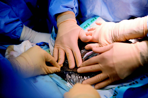
A suspicious lesion is generally removed to establish the diagnosis. If its site or size makes complete initial removal difficult, the specialist may choose targeted sampling. The specimen must allow assessment of possible invasion and, where possible, thickness: destroying a suspicious lesion with a cosmetic procedure can compromise this information.
After diagnosis, a second procedure, called wide local excision, may be needed around the original lesion site and scar. This does not necessarily mean the first operation was performed incorrectly: diagnostic biopsy and definitive treatment have different purposes. Even an initial report describing clear margins may still require an appropriate wider excision.
Before surgery, it helps to clarify which pathology report will guide the operation, whether the slides need review, and whether sentinel lymph node biopsy should be planned alongside wide excision. Patients should know when the final result will be available and who will discuss the next steps.
Sources for this section: American Academy of Dermatology — Melanoma clinical guideline; Cancer Research UK — Surgery to remove melanoma (reviewed 2025).
Surgical margins: interpreting the measurements
ESMO guidance specifies clinical margins of 0.5 cm for melanoma in situ, 1 cm for invasive melanomas up to 2 mm thick and 2 cm for thicker tumours. These are references for planning excision, not distances patients should measure themselves. The clinical margin marked on the skin and the microscopic margin reported by the pathologist are not interchangeable measurements.
Site, extent and histological type may require particular planning, especially on the face, at acral sites and in lentigo maligna. A 3 cm margin should not automatically be applied simply because a melanoma is thicker than 2 mm. The surgeon should explain the approach being used and how complete removal will be assessed.
If the final report identifies tumour at a margin, the case is reassessed to determine further management. This decision cannot be inferred from the visible scar’s length alone. Differences between international recommendations should be discussed in their clinical context rather than combining measurements from different schemes arbitrarily.
| Histological finding | Clinical margin |
|---|---|
| Melanoma in situ | 0.5 cm |
| Invasive, Breslow ≤ 2 mm | 1 cm |
| Invasive, Breslow > 2 mm | 2 cm |
Sources for this section: ESMO — Cutaneous melanoma: Clinical Practice Guideline (2024/2025).
Sentinel nodes and clinically involved lymph nodes
Sentinel lymph node biopsy looks for small melanoma deposits in the first nodes draining the tumour area when nodal involvement is not already clinically evident. Its indication depends on the pathology report and individual circumstances; it is not required for every melanoma. The dedicated child page explains selection, the procedure and interpretation of results.
A positive sentinel node does not automatically require removal of every node in that basin. The randomised MSLT-II trial found no melanoma-specific survival advantage from immediate completion dissection over ultrasound surveillance in the studied population; dissection improved regional control but increased lymphoedema. Surveillance must be organised and practically accessible.
Palpable nodes or nodes that appear suspicious on imaging represent a different situation from a microscopic deposit discovered in a sentinel node. They require diagnostic confirmation, staging and multidisciplinary discussion: the sequence of medical treatment and surgery should not be decided by automatically applying the sentinel-node pathway.
Sources for this section: Faries et al. — MSLT-II, NEJM (2017); NICE NG14 — Melanoma: assessment and management, recommendations.
Surgery and treatment before or after an operation
Adjuvant means after removal, aiming to reduce recurrence risk; neoadjuvant means before a planned operation. These terms are not interchangeable, and not every patient needs both. In higher-risk disease, decisions consider stage, general health, molecular characteristics where relevant, expected benefits and possible toxicities.
For some patients with resectable, macroscopic stage III melanoma, neoadjuvant immunotherapy is supported by randomised evidence, including NADINA. This does not justify extending it indiscriminately to thin or in-situ melanomas or every positive sentinel node. A response to treatment does not automatically mean surgery can be omitted.
References to interferon or cytokines in historical pages should not be interpreted as a list of today’s preferred options. Medical treatment has changed. Scientific evidence, regulatory authorisation and reimbursement are also separate matters: the treating centre verifies the indication and actual access in the country providing care.
Sources for this section: Blank et al. — NADINA, phase 3, NEJM (2024); National Cancer Institute — Melanoma Treatment (PDQ), health professional version.
Recurrence, metastases and local treatments
A new lesion on or near the scar, or along the lymphatic drainage pathway, needs assessment to distinguish recurrence, in-transit metastases and other conditions. Surgery may have a role when disease is limited and completely removable; when disease is widespread, repeated local excisions do not necessarily address the overall problem.
In metastatic melanoma, an operation may still be considered in selected circumstances to control a site or relieve symptoms, within a strategy that considers disease throughout the body. Surgery is not mandatory for every metastasis and does not remove the need for oncology assessment.
Isolated limb perfusion or infusion, electrochemotherapy and radiotherapy have specific indications in experienced centres. They are not equivalent alternatives to choose according to lesion size alone. Postoperative radiotherapy is not automatically indicated whenever several nodes are involved: potential local benefits and toxicity require individual discussion.
Sources for this section: National Cancer Institute — Melanoma Treatment (PDQ), health professional version; NICE NG14 — Melanoma: assessment and management, recommendations.
Lentigo maligna: surgery and selected alternatives
Lentigo maligna is a form of melanoma in situ; once invasive, it is termed lentigo maligna melanoma. This distinction changes treatment assessment. Abnormal melanocytes may extend beyond the visible edge and involve skin appendages: the colour seen with the naked eye does not reliably define the whole tumour.
Surgery remains the reference treatment. Staged excision or margin-controlled techniques, including Mohs surgery in appropriate settings, may be considered at delicate sites. Dermoscopy and other mapping tools may assist the team but do not replace histological assessment.
When surgery is impractical or would cause unacceptable morbidity, options such as radiotherapy or imiquimod may be considered for selected cases of in-situ lentigo maligna. Imiquimod requires specialist assessment, including consideration of off-label use. The pages on soft X-rays and laser explain the differences: inclusion of a technique in this index does not mean it is standard care or equivalent to excision.
Sources for this section: American Academy of Dermatology — Melanoma clinical guideline; NICE NG14 — Melanoma: assessment and management, recommendations.
Preparing, recovering and continuing follow-up
The wound may be closed directly or require a skin graft or flap, depending on the site and extent of removal. Before surgery, ask how anaesthesia, dressings, pain, daily activities and work will be managed. Report medications, allergies and relevant conditions without independently changing existing treatment.
At discharge, you should know how to contact the service and which signs warrant contacting it, such as persistent bleeding, increasing redness, fever or wound problems. Scars do not all heal at the same rate; your surgeon’s instructions take precedence over other patients’ experiences.
Wound review and oncological follow-up serve different purposes. Keep pathology reports, clarify the appointment schedule and continue skin surveillance. Completion of an operation alone cannot determine residual risk or justify stopping agreed follow-up.
Sources for this section: Cancer Research UK — Surgery to remove melanoma (reviewed 2025); Mayo Clinic — Sentinel node biopsy.
Experimental treatments: what is being studied
Research also concerns treatment around surgery. INTerpath-001 (NCT05933577) is a phase III study of an individualised neoantigen treatment, intismeran autogene/V940, combined with pembrolizumab after resection of high-risk melanoma. This is a therapeutic vaccine being studied in people who have already had cancer, not a preventive vaccine to offer the general population.
Another research direction examines how surgery and subsequent treatment might be adapted to the pathological response to immunotherapy: PRADO, with five-year findings published in 2026, contributes to this work. It should not be translated into a rule that tumour shrinkage makes surgery unnecessary. The phase Ib/II Morpheus-Melanoma study instead explores new immune-treatment combinations before surgery; that evidence level is not definitive confirmation for widespread use.
These examples describe research questions, not promises of cure or invitations to replace recommended care. Experimental treatment may be no better than standard care and may cause important adverse effects. Access requires eligibility criteria, informed consent and assessment at an authorised centre; location and availability must be checked in the registry and with the team. Registration of a study does not demonstrate efficacy, marketing authorisation or reimbursement. Do not stop or delay an agreed treatment to seek a trial independently.
Sources for this section: ClinicalTrials.gov — INTerpath-001, NCT05933577, phase 3; Hoeijmakers et al. — PRADO, 5-year follow-up, Nature Medicine (2026); Morpheus-Melanoma — Randomized phase 1b/2 study (2025); National Cancer Institute — Participate in Cancer Treatment Trials.
Sources and update
- National Cancer Institute — Melanoma Treatment (PDQ), health professional version
- American Academy of Dermatology — Melanoma clinical guideline
- Cancer Research UK — Surgery to remove melanoma (reviewed 2025)
- ESMO — Cutaneous melanoma: Clinical Practice Guideline (2024/2025)
- Faries et al. — MSLT-II, NEJM (2017)
- NICE NG14 — Melanoma: assessment and management, recommendations
- Blank et al. — NADINA, phase 3, NEJM (2024)
- Mayo Clinic — Sentinel node biopsy
- ClinicalTrials.gov — INTerpath-001, NCT05933577, phase 3
- Hoeijmakers et al. — PRADO, 5-year follow-up, Nature Medicine (2026)
- Morpheus-Melanoma — Randomized phase 1b/2 study (2025)
- National Cancer Institute — Participate in Cancer Treatment Trials
Editorial revision and source consultation: 17 October 2026. Study dates distinguish current evidence from historical documents. This date does not certify specialist clinical review.