What “superficial spreading” means
Superficial spreading melanoma, often abbreviated SSM, originates in melanocytes. It is the most frequent subtype in many fair-skinned populations. Reported proportions, often around two-thirds of cases, depend on the population and registration criteria rather than representing a universal percentage.
The name describes a growth pattern and does not guarantee that the tumour is shallow. A superficial spreading melanoma may be in situ or invasive. Understanding an individual case requires the histology report and, when present, measurement of the invasive component.
Compared with other common types of melanoma, it may have a recognisable phase of sideways spread, but its duration cannot be predicted by looking at the skin. There is no safe waiting period for a suspicious lesion.
Sites and relationship with moles
It can develop at different sites. The back and trunk in men and the legs in women are frequently described locations, but these patterns do not exclude other sites or groups. It can occur in adults, including older adults; an average age reported in a study is not a diagnostic threshold.
It may arise within a pre-existing mole, but many cases develop on skin where no mole was previously apparent. It should therefore not be regarded mainly as degeneration of a familiar mole. Individual susceptibility, personal or family history, mole count and UV exposure contribute to risk assessment.
Appearance and evolution
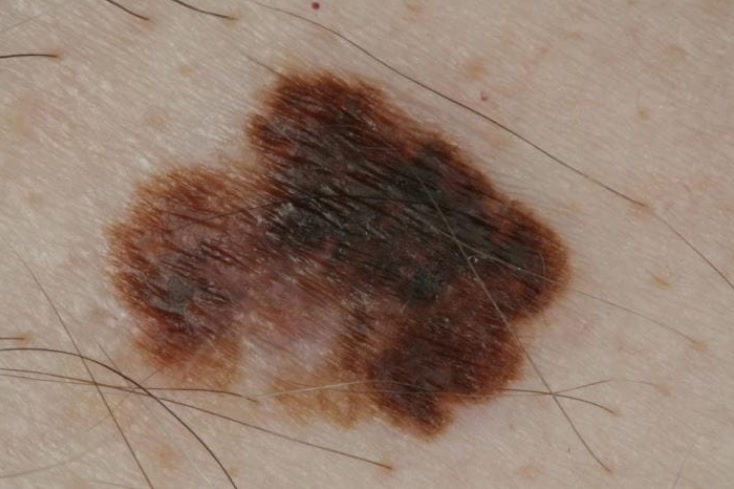
The lesion may be flat or slightly raised, asymmetric, with irregular, notched or blurred edges. Pigmentation can combine brown, black, blue, grey and pink with lighter or depigmented areas. These features do not always occur together, and some lesions are small.
During radial growth, cells extend sideways. Once they cross the boundary between epidermis and dermis, the melanoma is invasive. A new raised area or nodule within a patch can indicate an important change. Invasion may nevertheless already be present without a visible nodule.
The ABCDE rule helps track asymmetry, borders, colour, diameter and evolution. A lesion unlike the person’s other moles is also relevant. Ulceration, bleeding, persistent itching or rapid changes should be reported, but absence of symptoms does not exclude melanoma.
From examination to histological confirmation
Dermatological examination and dermoscopy guide suspicion. Physical examination alone does not confirm the diagnosis: tissue analysis is required. Where feasible, a suspicious lesion is removed completely for excisional biopsy. Location and size may require a different strategy selected by the specialist.
The report establishes whether melanoma is in situ or invasive. In invasive disease, Breslow thickness, ulceration and margins are key information. Sentinel lymph node biopsy and imaging are considered according to stage and risk rather than ordered automatically for everyone with this subtype.
Treatment and follow-up
Surgery is the main treatment for localised disease. A wider excision may be needed after diagnosis, with margins determined by the pathological findings. Additional treatment is discussed in selected higher-risk situations.
For unresectable or metastatic disease, options may include immunotherapy and targeted therapy when suitable molecular alterations are present. Radiotherapy and chemotherapy have specific indications; they are not interchangeable choices based simply on the subtype name. Our treatment guide introduces these pathways.
Prognosis depends mainly on disease extent and tumour characteristics. Follow-up is individualised after treatment. Skin self-examination and UV protection remain useful but do not replace appointments. Bring your report, treatment list and information about changes noticed between visits to the team.
Sources and update
- National Cancer Institute — Melanoma Treatment (PDQ)
- ESMO — Cutaneous melanoma: Clinical Practice Guideline (2025)
- DermNet — Superficial spreading melanoma
Editorial revision: 16 October 2026. This information does not replace medical assessment.