What it is and why prompt assessment matters
Nodular melanoma is an invasive melanoma characterised by relatively early growth into deeper skin. It is less frequent than superficial spreading melanoma. Estimates vary between populations and clinical series, so no single percentage describes every setting.
Rapid growth can lead to substantial thickness by diagnosis. This explains part of its clinical importance, but does not mean every nodular melanoma has already metastasised. Thickness, ulceration and spread to lymph nodes or other organs need individual assessment.
A pre-existing mole does not have to change: the tumour can appear as a new growth. A growing nodule warrants prompt assessment even when small, regular or painless.
Pigmented or amelanotic nodule
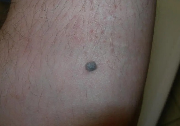
It may appear as a dome-shaped nodule or polypoid lesion, coloured black, blue or brown. Edges can be relatively regular and colour uniform; neither feature alone guarantees benignity.
Some lesions are pink, red or skin-coloured because they contain little visible pigment. This is an amelanotic melanoma presentation and may resemble a vascular or inflammatory lesion. Crusting, ulceration or bleeding can develop, but early lesions may have an intact surface and no symptoms.
Change from the original appearance matters. Recording when the bump appeared and how quickly it grew helps the clinician. An earlier photograph may be useful but should not delay assessment.
ABCDE and EFG: complementary reminders
The ABCDE rule is useful, but some nodular melanomas lack typical asymmetry or varied colours. EFG is an additional reminder for nodules: Elevated, Firm and Growing.
EFG is not a score that diagnoses cancer at home. Many benign growths are raised or firm. The combination of features and growth is what warrants attention. Do not wait for a lesion to reach 6 mm, bleed or become painful before requesting assessment.
A suspicious change between scheduled visits warrants earlier contact. Skin self-examination includes hard-to-see areas and more than familiar dark moles.
Diagnosis and look-alike lesions
Pigmented seborrhoeic keratosis, pigmented basal cell carcinoma, dermatofibroma and some vascular lesions can enter the differential diagnosis. External resemblance does not permit reliable distinction.
Dermoscopy may show residual pigment, structureless areas or atypical vessels. Absence of a pigment network does not exclude melanocytic origin. Histological examination is required to confirm the diagnosis and measure depth.
Complete excisional biopsy is performed where feasible. The report and clinical assessment guide subsequent investigations, including possible sentinel lymph node biopsy and imaging. A rapidly growing lesion should not simply be watched for a prolonged period without a specialist decision.
Treatment, prognosis and surveillance
Localised disease is treated surgically. Excision margins and other steps depend on the report and stage. Higher-risk or advanced disease may require immunotherapy or targeted treatment if molecular requirements are met. Radiotherapy and chemotherapy have selected roles.
The course cannot be predicted from the word “nodular” alone. Prompt diagnosis may allow treatment before spread, while greater thickness or metastases change the pathway. General statistics from different populations should not be applied directly to an individual.
After treatment, ask for a written follow-up plan and guidance on changes to report. The treatment and diagnosis guides can help prepare a discussion with the team. Surveillance includes the treated site and the whole skin because a separate new primary melanoma can also develop.
Sources and update
- National Cancer Institute — Melanoma Treatment (PDQ)
- ESMO — Cutaneous melanoma: Clinical Practice Guideline (2025)
- DermNet — Nodular melanoma
Editorial revision: 16 October 2026. This information does not replace medical assessment.