A site not readily visible from outside
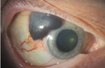
The ciliary body is part of the uvea behind the iris, contributing to aqueous humour production and focusing. Ciliary body melanoma may remain difficult to see early on and be recognised when it affects neighbouring structures.
It can cause visual problems, lens changes, increased eye pressure or inflammation. Pigmentary glaucoma and chronic uveitis are among conditions that can create diagnostic difficulties, but not everyone with these diseases has melanoma. An atypical presentation or unusual course may require further investigation.
Measuring the lesion and its extent
Ultrasound biomicroscopy assesses size, margins, shape and relationships with the iris, angle and sclera. It is combined with eye examination and other investigations when necessary. A photograph of the eye’s surface is insufficient to assess this location.
Assessment considers thickness, basal diameter, possible extraocular extension and available biological information. Thickness above 7 millimetres, mentioned in some historical descriptions, is not a universal threshold that alone determines metastasis or treatment. Risk reflects several factors and must be discussed in context.
Treatment options
Plaque brachytherapy and charged-particle radiotherapy may allow eye-preserving treatment in appropriate cases. Access to proton therapy depends on specialist centres and lesion characteristics. Fewer adverse effects cannot be promised for every tumour; proximity to structures needing preservation matters greatly.
Local resection is reserved for selected situations, considering the tumour base and surgical expertise. Extensive melanoma or a severely compromised eye may require enucleation. Planning should explain prospects for control, visual effects, complications and alternatives.
Advanced disease follows the uveal melanoma pathway rather than simply applying skin melanoma treatment. The eye melanoma page also explains systemic options and selection criteria.
Prognosis and surveillance
Sometimes delayed diagnosis and the characteristics of this site can be associated with higher risk, but they do not give everyone the same outlook. Size, extent and available pathological or molecular findings help personalise the discussion.
Follow-up checks local response, eye pressure, vision and complications alongside systemic surveillance set by the team. The liver is an important site in uveal melanoma follow-up. Report new symptoms rather than necessarily waiting for the next scheduled appointment.
Sources and update
Editorial revision: 17 October 2026. This information does not replace medical assessment. For very rare variants, some evidence comes from small series or case reports.