Nevoid melanoma, first proposed by Schmoeckel et al, is one of a diverse group of melanomas that have histological features similar to those of a benign nevus, also known as a “nevus”. Nevoid melanoma usually appears as an approximately 1 cm brown nodule on the trunk or proximal regions of young adults, with a higher incidence in males about age 43.
This type of melanoma initially develops from an existing melanocytic nevus, a benign lesion of the skin. Although most melanocytic nevi do not develop into cancer, some can develop into melanoma. An estimated 20% to 30% of melanomas result from a pre-existing nevus. However, most melanomas develop on previously normal skin, rather than from a nevus.
Like other types of melanoma, nevoid melanoma is caused by an uncontrolled growth of cells that produce melanin, the pigment that gives skin color. This uncontrolled growth can lead to the formation of malignant tumors.
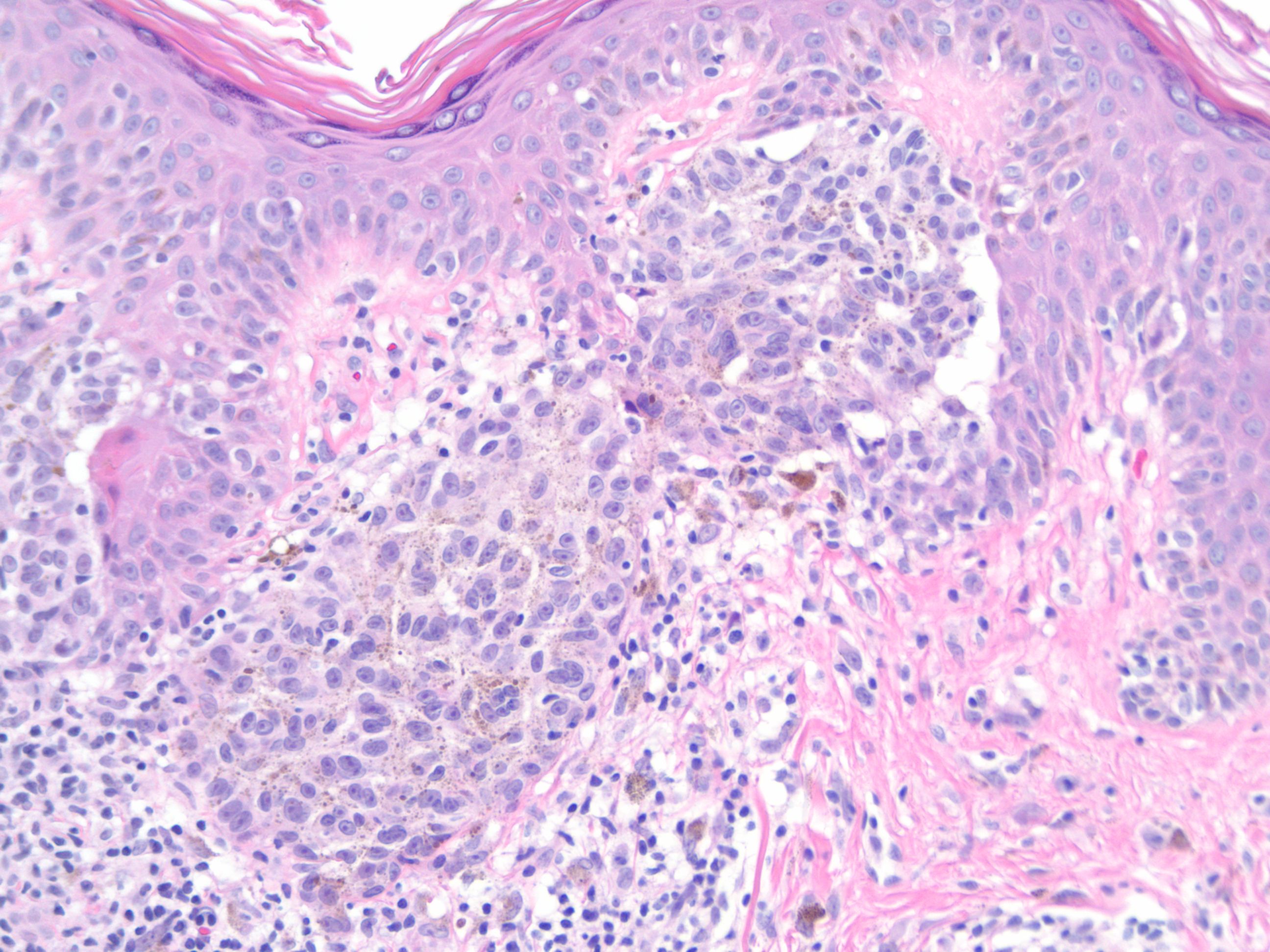
Histologically, nevoid melanoma has a verrucoid or domed pattern. The lesion is well circumscribed laterally, while the fundus is poorly defined. Junctional activity is rarely observed, while mitoses are present. There are nests of epithelioid melanocytes. There is a risk of confusing it with a dermal nevus.
Nevoid melanoma can manifest as a pigmented lesion with a variety of colors, including black, brown, blue, red, or white. Changes in the size, shape or color of an existing mole, or the appearance of a new mole with these characteristics, may be signs of melanoma.
Metastases and local recurrences after excision are described in the case of nevoid melanoma. Consequently, it is essential to consult a dermatologist if you notice changes in a pre-existing nevus or the appearance of a new nevus with these characteristics. Early diagnosis of melanoma can significantly improve the chances of effective treatment.