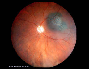
Melanoma of the eye is the most common of the cancers of the eye, the cancer originates from the melanocytes present in the eye. According to data from the National Health Service (NHS), UK about 500 new cases of ocular melanoma are diagnosed in Great Britain, the highest incidence being around 50 years of age. Approximately 2,350 new cases of eye cancer were diagnosed in the United States in 2009, most of which were melanoma. The incidence has grown slowly over the past 25 years. Treatment is effective if the tumor is discovered in the early stages and 85% survive for at least 5 years (Mayo Clinic data). The symptoms are variable: a dark spot in the iris, irregular vision, loss of peripheral vision in one eye, pain, flashes of light, etc. The risk factors include light eyes, chromosomal abnormalities (chromosome 3) , dysplastic nevus syndrome and excessive exposure of the eye to ultraviolet rays. Diagnosis is made by direct and/or indirect ophthalmoscopy, ultrasound, fluorescence angiography, CT scan, MRI, biopsy. In the following chapters we will delve into the topics of melanoma of the iris, choroid and ciliary body.
Therapy
Radiation therapy recommended in small or medium-sized tumors:
- Plaque radiotherapy: it is the most common therapy for posterior ocular tumors (choroidal and ciliary body) and consists in suturing a small radioactive metal object to the ocular wall adjacent to the tumor, once the tumor has been destroyed the plaque is removed, sometimes the patient must remain in hospital for the entire duration of treatment.
- Proton Beam radiotherapy: small rings of tantalum are placed at the edge of the tumor in order to direct the radioactive particles towards the tumor.
- Stereotactic radiotherapy: concentrates the radiation dose to the melanoma in small fractions.
Types of surgery:
- Enucleation: after surgery an artificial eye must be applied in the orbit and is performed in case of extensive tumors.
- Iridectomy: in tumors of the iris.
- Iridocyclectomy: removal of the iris and the adjacent ciliary body.
- Trans scleral local resection: the tumor is eliminated by making an opening in the wall of the eye, this technique is used in large tumors by associating it with plaque radiotherapy to reduce recurrences.
- Trans-retinal endoresection: The melanoma is removed through the retina and is performed when the tumor is close to the optic nerve. The laser is used to reduce recurrences.
- Transpupillary thermotherapy: laser therapy that heats the tumor using infrared laser radiation. It lasts about 30 minutes under local anesthesia and is used for small melanomas or when there is an uncertain diagnosis between malignant melanoma and benign nevus.
- Intraocular injections: treatment under local anesthesia and steroids or angiogenic factors are injected,