Primary or secondary: the first distinction
Melanoma involving the central nervous system is much more often metastatic than primary. A brain metastasis comes from melanoma originating elsewhere; primary CNS melanoma arises from melanocytic cells within nervous system structures, particularly the meninges. Finding a pigmented brain mass alone does not establish primary origin.
Primary meningeal melanocytic tumours include circumscribed and diffuse lesions with different behaviour. A melanocytoma is not automatically melanoma. Reports at sites such as the pineal region are exceptional and do not justify a universal estimate of 1% of all melanomas.
Symptoms and investigations
Presentation depends on location. Persistent headache, seizures, cranial nerve problems, changes in strength or sensation, imbalance and spinal signs may require investigation. Obstruction of cerebrospinal fluid pathways can cause hydrocephalus. Haemorrhage can occur, but not every neurological symptom indicates melanoma.
MRI with appropriate protocols examines the brain and, when indicated, spine and meninges. Searching for a primary site outside the CNS, including skin and eye, is part of establishing origin. Histology, immunohistochemistry and sometimes molecular testing complete assessment; imaging alone cannot define all aspects of biological aggressiveness.
Seizures, sudden deficits or a severe acute headache require urgent care. Persistent symptoms should be discussed with a clinician rather than independently attributed to cancer.
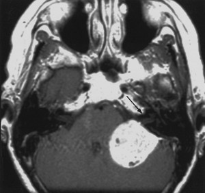
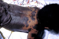
Neurocutaneous melanocytosis is not synonymous with melanoma
Neurocutaneous melanocytosis, also called neurocutaneous melanosis, combines congenital melanocytic nevi with melanocytic proliferations in the nervous system. Neurological abnormalities and, in some people, malignant transformation can occur. Melanocytes in the CNS do not, however, mean melanoma is already present.
It is generally a sporadic developmental condition associated with somatic mosaicism, often involving NRAS alterations, rather than a common inherited disorder following a simple familial pattern. A historical reference to “fewer than 200,000 people” is not a reliable estimate of its prevalence.
Large, giant or multiple nevi and neurological findings guide specialist paediatric assessment. Hydrocephalus, epilepsy and cranial or spinal problems are possible complications. MRI indications and surveillance depend on the phenotype; they should not be applied indiscriminately to every child with a small mole.
Treatment and evidence limitations
For brain metastases, surgery, stereotactic radiotherapy and systemic treatments may be considered according to lesion number and size, symptoms and disease outside the CNS. It is incorrect to describe all spinal involvement as having no treatment options. Goals may include disease control, protection of function and symptom relief.
Corticosteroids may reduce symptomatic swelling when indicated but do not treat the melanoma itself and require medical management. Decisions must also consider possible immunotherapy. Exceptionally rare primary tumours require planning according to resectability and integrated diagnosis. Radiotherapy and systemic strategies are discussed at expert centres, often using evidence limited to small series.
Rehabilitation and supportive care may accompany cancer treatment. Ask whether the diagnosis is primary or metastatic, which goals are realistic and which symptoms require immediate contact.
Sources and update
- NCI — Melanoma Treatment (PDQ)
- EURACAN — Primary meningeal melanocytic tumours (2024)
- Kinsler et al. — NRAS mosaicism and neurocutaneous melanosis
Editorial revision: 17 October 2026. This information does not replace medical assessment. For very rare variants, some evidence comes from small series or case reports.